
| Start of section
Production, amateur Radio amateurs Aircraft model, rocket-model Useful, entertaining |
Stealth Master
Electronics Physics Technologies Inventions |
Secrets of the cosmos
Secrets of the Earth Secrets of the Ocean Tricks Map of section |
  |
| Use of the site materials is allowed subject to the link (for websites - hyperlinks) | |||
Navigation: => |
Home / Patent catalog / Catalog section / Back / |
|
INVENTION
Patent of the Russian Federation RU2146883
![]()
METHOD FOR TREATMENT OF ACUTE OBSTURATION CHOLECYSTITIS
The name of the inventor: Evgeny Pavlovich Kuznetsov
The name of the patent holder: Kuznetsov Evgeniy Pavlovich
Address for correspondence: 426069, Udmurt Republic, Izhevsk, ul.Pesochnaya, 22, kv.76, Kuznetsov Evgeniy Pavlovich
The effective date of the patent: 1997.02.25
The invention relates to the field of medicine, in particular to surgery. Under the control of ultrasound scanning, the gallbladder is punctured through the place of the liver capsule passage into the serous cover of the bottom of the gallbladder. As a sanitizing agent, 0.8% NaClO solution is used. The needle is advanced to the area of the neck of the gallbladder. Change the direction of the needle. Control its position by ultrasonic scanning throughout the entire sanation. Achieve the state of an optically transparent medium in the lumen of the gallbladder. Through the same puncture needle, a monophilic light guide is inserted. Laser irradiation of the cavity of the gallbladder with NaClO solution is carried out as a light-conducting and light-scattering medium. The method allows to increase the effectiveness of conservative treatment, to refuse urgent or urgent surgical interventions in patients with low tolerance to the latter and to perform delayed or planned interventions.
DESCRIPTION OF THE INVENTION
The invention relates to the field of medicine, namely surgery, and is used in the treatment of acute obstructive cholecystitis in patients with low tolerance to radical surgery-a high risk of surgical intervention and a high anesthetic risk.
Known is a method for treating inflammatory diseases of the bile ducts, consisting in that through the drainage installed in the holedoch during the surgical intervention, the laser guide of the laser therapy unit is inserted in the postoperative period and endobiliary irradiation is performed (Mokeev AF Laser application in the treatment of bile duct obstruction benign - in the journal "Herald of Surgery", -1986, -N 11, -s. 11-15).
However, this method does not have sufficient efficiency, because Bile, located in the lumen of the bile ducts with cholangitis, absorbs and dispels a significant part of the laser radiation with particles of pigment suspension, detritus and fibrin flakes.
A method for treating acute obstructive cholecystitis is known, including puncture of the gallbladder percutaneously transhepatic under the control of ultrasound scanning, emptying the gallbladder and introducing antibiotics into its cavity at high concentrations (see Krylov, NV, Vyazitsky, PO, Seleznev, Yu.K. Etc. Controlled decompression of the gallbladder in acute cholecystitis - in the journal "Surgery", -1986, -N 2, -p. 60-64). The implementation of transhepatic puncture was justified by the need to restore the germicity of the cavity of the gallbladder, achieved by the obstruction of the puncture channel with liver tissue. Antibiotic was introduced to suppress pathogenic microflora in the cavity of the gallbladder.
However, the known method, taken as a prototype, in the presence of pronounced inflammatory-destructive changes in the wall of the gallbladder, causing the fragility of tissues in the area of the gallbladder bed, has the disadvantages that when the needle moves, the gallbladder wall can split off from its bed With the formation of hematomas and abscesses. In addition, with transhepatic access it is impossible to reach the area of the Hartmann pocket with the needle, where the largest amount of detritus is accumulated and thereby ensure the complete sanation of the gallbladder cavity without extraction of the needle or additional trauma to the liver parenchyma. Thus, in the cavity of the gall bladder there are areas where sanation is difficult, and the use of a solution of antibiotics as a sanitizing agent in some cases is ineffective in the resistance of microflora to it or inapplicable in the case of an intolerant antibiotic patient
The essence of the claimed invention lies in the fact that according to the method of treatment of acute obstructive cholecystitis, including puncture of the gallbladder under the control of ultrasonic scanning and sanitation of its cavity through a puncture needle, the insertion of the puncture needle is carried out along a path passing through the place of passage of the liver capsule into the serous cover of the bottom of the gallbladder , As a sanitizing agent, 0.8% NaClO solution is used, the needle is advanced to the area of the neck of the gallbladder, changing the direction of the needle and controlling its position by ultrasonic scanning throughout the entire sanation, and after reaching an optically transparent medium in the gallbladder lumen, laser irradiation of its cavity Through a monophilic light guide introduced through the same puncture needle, using a solution of NaClO as a light-conducting and light-scattering medium.
The use of the claimed method significantly increases the effectiveness of conservative treatment, allowing to refuse urgent or urgent surgical interventions in patients with low tolerance to the latter and to perform delayed or planned interventions.
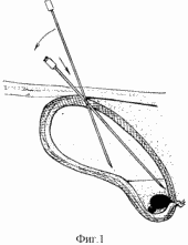 |
The claimed method uses access through the site of the liver capsule transfer to the serous cover of the bottom of the gallbladder. In acute cholecystitis, the wall of the gallbladder is thickened, impregnated with fibrinous exudate, so when the gallbladder is emptied and simultaneously disconnected from the biliary tree, this wall provides a reliable obstruction of the puncture channel after the extraction of the needle and excludes leakage of bile into the abdominal cavity. Better channel obturation is also facilitated by its direction tangential to the wall of the gallbladder (see Figure 1). With this access, it is possible to change the position of the needle during manipulation without the risk of injury to the liver parenchyma or the gallbladder wall detachment from the bed, which allows for a more complete sanation of the areas of the gallbladder that are difficult to access in other techniques. The use of a solution of NaClO as a sanitizing agent in a concentration of 0.8% makes it possible to effectively control any microflora, incl. Anaerobic. This antiseptic is a powerful oxidizer. In addition, unlike other oxidizing antiseptics containing peroxide compounds, when interacting with pus and detritus in NaClO solution, microbubbles of gas do not form, preventing ultrasound observation of the manipulation, which allows to control the process of sanation of the cavity of the gall bladder throughout the entire manipulation. |
The ability to change the direction of the needle in the cavity of the gallbladder and move it to the neck area allows to achieve mechanical cleansing of the gallbladder walls by flushing detritus with a jet of antiseptic injected under pressure from the cut.
Thus, with the use of this access and antiseptic, it is possible to achieve a much greater completeness of sanation of the gallbladder cavity, to achieve cleansing of the surface of the gallbladder mucosa and optical uniformity and transparency of the contents of the gallbladder, i.e. Create conditions for the performance of laser irradiation of its cavity. After the optical transparency of the contents of the gallbladder is reached, 20-30 ml of the antiseptic solution is left in its cavity, which is used as a light-conducting medium. The absence of detritus on the surface of the mucosal coating reduces the absorption and loss of energy of laser radiation. In addition, laser radiation, in addition to its photobiological action, triggers a series of photochemical reactions, generally enhancing the antibacterial activity of the NaClO solution.
The possibility of moving the needle under the control of ultrasound scanning to the region of the neck of the gallbladder allows to irradiate the obturation zone and, through the wall of the gall bladder, the adjacent tissues, vascular and duct structures, which leads to a decrease in the edema zone, better microcirculation in the wall of the gallbladder, Duct, the surrounding cellular spaces, which contributes to a more rapid recovery of the patency of the cystic duct, and enhances the effect of proper decompression.
The method is carried out as follows. Preliminary performed ultrasound examination of the gallbladder and surrounding tissues, which assess the anatomical features of the location of the gallbladder, assess the severity of inflammatory-destructive changes in its wall, changes in organs and tissues adjacent to the gallbladder.
The condition of the puncture of the gallbladder is thickening as a result of the inflammatory edema of its wall more than 0.6 cm, the absence of signs of the presence of fluid in the subhepatic space and signs of peritonitis, the absence of bile hypertension, estimated by the diameter of the choledocha less than 0.8 cm. The presence of obturation cholecystitis is confirmed by being in Lumen gallbladder concrements, its increase, tension, pain in its projection, the presence of signs of intoxication, thickening its walls.
Then, under the control of ultrasound scanning, a percutaneous puncture of the gallbladder with a needle 1.0-1.3 mm in diameter is made through the place of the liver capsule passage into the peritoneal cover of the gallbladder floor. The length of the needle should allow it to reach the point of the Hartmann pocket of the gallbladder from the puncture point.
After the needle enters the cavity of the gallbladder, aspiration is carried out through the lumen of the needle of its contents. At the same time, as the gallbladder is emptied, the needle under the ultrasound scanning is advanced, changing the mutual position of the needle and the sensor, to the sites of the detritus accumulation.
After possibly more complete emptying of the gallbladder, sanitation of its cavity is performed with a 0.8% NaClO solution and under the control of ultrasound scanning. The injection and removal of the sanitizing solution is carried out through the lumen of the puncture needle, the position of which, as far as the sanation is concerned, is changed to more completely remove detritus from the neck region and the Hartmann pocket of the gallbladder. In this case, the jet of the antiseptic solution delivered under pressure promotes the mechanical removal of the fibrin deposit, pigment suspension and mucus from the gallbladder walls, and the oxidizing properties of the NaClO solution contribute to the effective sanitation and destruction of the pathogenic flora. Sanitation is performed until an optically transparent medium is reached in the lumen of the gallbladder, which is controlled by the nature of the aspirate coming from the needle and by the disappearance of the accumulations of the hyperechoic suspension according to ultrasound scanning data.
Changing the position of the needle, its working cut is moved to the region of the neck of the gallbladder, aspirate the solution of NaClO contained in the cavity of the gallbladder so that 20-30 ml remain in the lumen. Then, through the lumen of the puncture needle, a monophilic light guide is connected to the region of the neck of the gallbladder, connected to a laser emitter of low power. Perform the irradiation of the cavity of the gallbladder for 5-10 minutes, depending on the power and wavelength of the radiation.
The light guide is then removed with the needle. In the absence of negative dynamics, the procedure is performed once, with a repeated attack - up to 3 times.
Example 1 . Patient E., 77 years old, entered the on-duty surgical clinic with the diagnosis: cholelithiasis, acute calculous cholecystitis, atherosclerotic cerebrosclerosis, coronary heart disease, postinfarction cardiosclerosis, hypertension, circulatory insufficiency 2A.
At admission, the condition is of medium severity, subfebrile temperature, signs of intoxication are expressed, symptoms of irritation of the peritoneum are not present.
From laboratory data: high leukocytosis, shift of the leukocyte formula to the left.
Upon admission to the ultrasound study, a gallbladder measuring 12.6 × 4.3 cm, enlarged, strained, the wall unevenly thickened, laminated to 0.8 cm in thickness. In the area of the bed, an anechogenous, irregularly shaped formation is formed, suspicious for an abscess, measuring up to 1.2 x 0.7 cm, with an indistinct contour. In the lumen of the gallbladder - more than 5 concrements 0.5 - 0.9 cm in diameter located in the area of the Hartmann pocket, with a clear acoustic shadow. Ibid - the level of hyperechoic suspension. Holedoch up to 0.8 cm in diameter, widened slightly. Conclusion: cholelithiasis, acute obturation calculous cholecystitis. Forming abscess of the area of the gallbladder bed.
Infusion therapy, antispasmodics, anticholinergics, analgesics, antibiotics, cardiac glycosides, hypotensive agents are prescribed. Within 12 hours the attack is not stopped, pains remain, the bottom of the gallbladder is palpated, intoxication is accumulating, there are no symptoms of irritation of the peritoneum. 12 hours after admission, under local anesthesia, under the control of ultrasound scanning, a percutaneous puncture of the gallbladder was performed along a trajectory passing through the place of passage of the liver capsule into the serous cover of the bottom of the gallbladder with a needle 1.2 mm in diameter. Removed up to 130 ml of viscous purulent bile with pigment suspension. As the gallbladder is uncovered, the needle is advanced to the area of the Hartmann pocket, for which the angle of inclination of the needle relative to the ultrasound scanner sensor is increased. 35 ml of thick purulent bile with putty pigment suspension was removed from the area of Hartmann's pocket. A 0.8% NaClO solution was sanitized, for which the antiseptic solution was pumped through the puncture needle into the gallbladder lumen in 25-50 ml portions and then aspirated through the same needle with a syringe. At the same time, ultrasound monitoring of the position of the needle tip in the lumen of the gallbladder was performed. When a sanitizing solution enters the gallbladder lumen with ultrasound scanning, a short-term appearance of intense heterogeneity of the contents is noted, associated with the mixing of the sanitizing solution with detritus and pus.
270 ml of the sanitizing solution were used to obtain a transparent aspirate, after which 80 ml of the contents were removed from the gallbladder cavity so that about 30-40 ml of the sanitizing solution remained in the gallbladder lumen (the diameter of the gallbladder decreased to 2.4 cm); The puncture needle is advanced to the neck of the gallbladder, a quartz monophilic light guide with a diameter of 0.6 mm is passed through its lumen, connected to a laser therapy apparatus based on a helium-neon laser LG-75 with a power at the end of the optical fiber of 5.0 mW. Irradiation of the cavity of the gallbladder with an exposure of 8 min was performed, after which the light guide was removed together with the needle.
Immediately after performing the manipulations, the pains significantly decreased. Dyspeptic phenomena were stopped after 16 hours. Normalization of the temperature occurred after 24 hours. Normalization of the leukoformula was achieved after 2 days.
In the ultrasound examination on the 7th day the gallbladder measuring 7.4x3.1 cm was not enlarged, atonic, deformed in the region of the bottom and the cervix, the wall of uneven thickness to 0.6 cm, thickened mainly in the region of the bed of the gallbladder, homogeneous, Sharply increased echogenicity. In the lumen of the gallbladder - a homogeneous liquid content and several concrements with a clear acoustic shadow with a diameter of up to 0.9 cm, located in the area of the body of the gallbladder.
The patient is discharged for outpatient treatment on the 7th day.
Example 2 . Patient N., 72 years old, entered the surgical clinic on duty with a diagnosis: cholelithiasis, acute calculous cholecystitis. Concomitant pathology is represented by atherosclerotic coronary heart disease, ischemic heart disease, obstructive chronic bronchitis, pulmonary emphysema, respiratory failure, common osteochondrosis with radicular syndrome; 6 months ago has transferred or carried operation in occasion of an adenoma of a prostate, functioning tsistostoma.
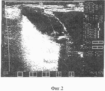 |
At admission, the condition is severe, severe intoxication, signs of dehydration; From laboratory data - high leukocytosis with a shift of the leukocyte formula to the left, an increase in the level of bilirubin, transaminases and residual blood nitrogen. In ultrasound examination, the gallbladder is 10.9 × 5.7 cm in size, enlarged, strained, its wall sharply thickened to 1.1 cm, layered, in the lumen of the gallbladder - a single calcaneus wedged into the neck, 1.9 cm in diameter (see Fig. 2). No signs of the presence of fluid in the subhepatic and near-bubble space were found. Holedoch is not enlarged, 0.6 cm in diameter. Conclusion: LCB, acute calculous cholecystitis. |
Spasmolytics, infusion therapy, antibiotics, cholinolytics, analgesics are prescribed. 4 hours after the patient's admission, he underwent local anesthesia and ultrasound control of the puncture of the gallbladder with a needle 1.2 mm in diameter along a trajectory passing through the place where the liver capsule passed into the serous cover of the bottom of the gallbladder. After the needle hit the cavity of the gallbladder, 120 ml of bile mixed with purulent hemorrhagic exudate was removed from the latter. The sanation of the gallbladder cavity with a NaClO solution of 0.8% was started: the sanitizing solution was administered in portions of 40 ml through a puncture needle with subsequent aspiration. During the sanitation, the needle was moved, while changing its inclination relative to the sensor, until the tip of the needle reached the area of Hartmann's pocket. During the sanation, 200 ml of sanitizing solution was used to obtain a transparent aspirate from the cavity of the gallbladder. Then, after removing 50 ml of contents from the cavity of the gallbladder so that its diameter decreased to 2.9 cm, a monophilic light guide with a diameter of 0.6 mm was connected through the same puncture needle to a laser device based on the LG-75 emitter with a power of The end face of the optical fiber is 5.0 mW. Its position is controlled by ultrasonic scanning. Irradiation of the gallbladder cavity was performed for 8 min. After that, the light guide is taken out together with the puncture needle.
The pain subsided immediately after the completion of the manipulation. Dyspeptic phenomena are stopped and within 2 hours after the manipulation.
In a day the pains arose again. With ultrasound examination, the gallbladder is 9,5x4,7 cm in size, enlarged, strained, the wall is thickened to 1.5 cm, layered, of uneven thickness, in the lumen of the gallbladder - a concrement up to 1.9 cm in diameter located in the neck region. The contents of the gallbladder are sharply heterogeneous. There were no signs of the presence of fluid in the subhepatic space.
Under local anesthesia and ultrasound control, a puncture of the gallbladder was repeated with a 1.2 mm needle along the same trajectory. From the lumen of the gallbladder, 100 ml of light opalescent exudate with strong chlorine odor without bile impurities are removed. The sanation of the cavity of the gallbladder and laser irradiation of the cavity of the gallbladder according to the above described procedure is similar to the first manipulation.
The pain subsided immediately after the completion of the manipulation; In the subsequent pain was not resumed. Normalization of indicators of the number of blood leukocytes and the leukocyte blood formula was achieved on the 5th day.
On the 7th day, with an ultrasound examination, a gallbladder measuring 6.9x3.3 cm, deformed by two constrictions in the body region, the wall of the gall bladder of uneven thickness, up to 0.7 cm. The calculus freely moves in the lumen of the gallbladder when the patient's position changes.
The patient was transferred to the therapeutic department for the treatment of concomitant diseases on the 9th day.
CLAIM
A method for the treatment of acute obstructive cholecystitis, comprising puncturing a gallbladder under the control of an ultrasound scan and sanitizing its cavity through a puncture needle, characterized in that the insertion of a puncture needle is performed along a path passing through the place of the liver capsule passage into the serous cover of the bottom of the gallbladder, Use a 0.8% solution of NaClO, the needle is advanced to the region of the neck of the gallbladder, changing the direction of the needle and controlling its position by ultrasonic scanning throughout the entire sanation, and after reaching an optically transparent medium in the gallbladder lumen, laser irradiation of its cavity through a monophilic light guide , Introduced through the same puncture needle, using a solution of NaClO as a light-conducting and light-scattering medium.
print version
Date of publication 28.01.2007gg
![]()

Comments
When commenting on, remember that the content and tone of your message can hurt the feelings of real people, show respect and tolerance to your interlocutors even if you do not share their opinion, your behavior in the conditions of freedom of expression and anonymity provided by the Internet, changes Not only virtual, but also the real world. All comments are hidden from the index, spam is controlled.