|
Start of section
Production, amateur Radio amateurs Aircraft model, rocket-model Useful, entertaining |
Stealth Master
Electronics Physics Technologies Inventions |
Secrets of the cosmos
Secrets of the Earth Secrets of the Ocean Tricks Map of section |
|
| Use of the site materials is allowed subject to the link (for websites - hyperlinks) | |||
Navigation: => |
Home / Patent catalog / Catalog section / Back / |
|
INVENTION
Patent of the Russian Federation RU2199957
![]()
COMPLEX METHOD OF DIAGNOSTICS AND SURGICAL TREATMENT OF ACUTE CHOLECYSTITE IN PATIENTS WITH HIGH OPERATIONAL ANESTHESIA RISK
The name of the inventor: Suzdaltsev IV; Arkhipov O.I.
The name of the patentee: Suzdaltsev Igor Vladimirovich
Address for correspondence: 355017, Stavropol, ul. Mira, 310, Medakademiya, Patent Department
Date of commencement of the patent: 2000.08.10
The invention relates to medicine, surgery, can be used to diagnose and treat acute cholecystitis in patients with high operational anesthesia risk. Perform ultrasound examinations, chromoduodenoscopy. Consider the time of chromoduodenoscopy of the secretion of colored bile from the fat nipple. Perform cholecystostomy under local anesthesia with a block of bladder duct with the imposition of a single suture suture. Control the state of cholecystostomy using methylene blue. In the production of the green separated by safety drainage after the administration of methylene blue, chemical mucoclasis is performed. Use medicamentous drugs to reduce edema and spasm of the fat nipple. The method allows to reduce postoperative complications.
DESCRIPTION OF THE INVENTION
The invention relates to the field of medicine, namely, to surgery.
Acute cholecystitis in recent decades has become a social problem, with 34 - 40% of the total number of patients entering treatment facilities with this pathology, are elderly and old people [1].
According to modern ideas, in the pathogenesis of acute cholecystitis in this category of patients, in addition to acute obturation of the cystic duct leading to a sharp increase in intravesical pressure and hyperextension of the gallbladder wall, great importance is attached to the vascular factor [2].
Disturbance of hemocirculation in the wall of the gallbladder in connection with sclerosis and vascular thrombosis causes a rapid (within a few hours after the disease) development of destructive forms of acute cholecystitis [3, 4].
However, in patients older than 60 years, despite the high percentage of destructive forms, the rapid course of the disease with a pronounced clinical picture from the side of the abdominal cavity is either absent or does not last long. Pathomorphological changes do not have time to be realized in symptoms. After that, the phenomena of intoxication against the background of existing age-related changes and concomitant pathology are on the foreground in the development of acute cholecystitis [5, 1]. Thus, the operation undertaken to "change the clinical picture" from the abdominal cavity, is belated. As a result, postoperative mortality remains stably high, amounting to more than 20% [1].
In the light of the above, the significance of additional instrumental methods for diagnosing the bladder duct block becomes clear.
At present, the following methods of diagnostics have found the most widespread:
1. Ultrasound examination (ultrasound). The principle of operation of diagnostic instruments is as follows: the object is subjected to a directional ultrasonic beam and echoes reflected from the boundary of two media with different acoustic densities are recorded [6]. Simplicity, safety, the possibility of examining the patient an unlimited number of times, regardless of the severity of his condition makes the ultrasound study irreplaceable in the diagnosis of acute cholecystitis.
2. Duodenal sounding [1, 7]. It was proposed in 1919. There are three portions of bile: "A" - the contents of the duodenum (golden yellow liquid), "B" - cystic bile (dark olive color, viscous liquid), "C" - hepatic bile (less viscous, transparent, golden liquid) . To stimulate the bubble reflux, magnesium sulfate is introduced through the probe into the duodenum. The absence of portion "B" indicates the block of the cystic duct.
3. A sample of D. Febres. For a clearer definition of gallbladder D. Febres (1942) [8] proposed a test with methylene blue. He found that methylene blue, injected inside (in capsules), is excreted partly by the liver, partly by the kidneys. When excreted from the liver, it turns into a colorless leukain, which again turns into a chromogen in the gallbladder and stains the bladder bile in blue-green color, while the bile of portions "A" and "C" containing the leuco base is colored in a normal color. The intensity of the color depends on the concentrating ability of the gallbladder mucosa, and on the pH of the bile, a decrease in the latter helps oxidize the dye and stain the bile. The shift of the pH of the bile to the acidic side indicates an inflammatory process in the gallbladder. In patients with acute calculous cholecystitis, the pH of the gall bladder averages 5.3 compared with 6.9 in the control group [9].
Thus, using the D. Febres test with duodenal sounding, it is easy to distinguish one portion from another, since only the gallbladder is colored in blue-green (malachite) color.
4. Accelerated chromatic duodenal sounding [10]. It is a combination of duodenal sounding and D. Febres. When performing this study, 0.4% solution of indigo carmine is used intravenously instead of methylene blue, which gives a similar effect. The patient is injected with a probe into the duodenum and then 5 ml of a 0.4% solution of indigo carmene. To stimulate the bubble reflux, 40-50 ml of 25% solution of magnesium sulphate is used. The coloration of the gall bladder, obtained by sounding, excluded acute cholecystitis, since it indicated the permeability of the cystic duct.
The listed types of research are not without a number of shortcomings.
With ultrasound, the information in the diagnosis of acute cholecystitis reaches 92-98% [11, 12]. In some cases, both false-positive and false-negative results are possible. The reason for them is obvious in the presence of gaseous gut (reflex paresis of the gastrointestinal tract) and, in this connection, the impossibility of a clear visualization of the gallbladder.
Different types of duodenal studies are not without flaws. When duodenal sounding is difficult to judge the patency of the cystic duct, since the color difference between the vesicle and hepatic bile is a rather subjective criterion. The D. Febres sample allows more accurate determination of the gallbladder, but since methylene blue is administered per os, the informative value of the method is reduced in the presence of diseases of the gastrointestinal tract.
Accelerated chromatic duodenal sounding has its negative aspects. These include: 1. Difficulties, and sometimes the impossibility of carrying out duodenal sounding in patients with acute cholecystitis due to frequent vomiting, motor trouble, duodenosis, and throwing the contents of the duodenum into the stomach. Failures, according to our data, account for 60%. 2. Duration of the sounding procedure. 3. Absence of reliable criteria for finding the probe in the duodenum. 4. Absence of colored bile when using this technique does not always indicate blockade of the cystic duct - cases of choledoch obstruction are possible.
The prototype of our proposed study is chromoduodenoscopy (HDS) - a visual observation of the isolation of paint from the large nipple of the duodenum (BSDK) with the help of a duodenoscope [13]. HDS, as well as accelerated chromatic duodenal sounding, is based on a sample of D. Febres.
The procedure is as follows: 1) preparation - on an empty stomach and, if necessary, in case of an emergency CDS, gastric lavage; 2) Premedication - 30 minutes prior to the study, the administration of promedol, atropine sulfate, diphenhydramine and, if necessary, aeron under the tongue; 3) intravenous injection of 5 ml of 0.4% solution of indigocarmine for 10-15 minutes before the study; 4) the introduction of an endoscope under local anesthesia with a 2% solution of dicain; 5) examination of the esophagus, stomach, duodenum and BSDK. The authors did not cause a vesicle reflux with magnesium sulfate. According to the intensity of the color of the extracted bile and dye, in the opinion of the authors, it is possible to judge with certainty the function of the gallbladder, the degree of obstruction of the cystic duct, the presence of dilated choledoch and the violation of motility of the biliary tract. By the duration of the interval between the periods of sphincter contraction, Oddy is judged on his functional and organic changes. If there is a violation of the motility of the bile ducts and the dilated choledocha of a functional nature, the interval between the ejections of the colored bile increases 2-3 times in comparison with the norm.
The described method has several disadvantages: 1. Preparations used for premedication (promedol, atropine sulfate, diphenhydramine) affect the contractility of the gallbladder and sphincter apparatus [14], which can lead to artifacts. 2. There is no stimulation of cystic reflux, which does not allow one to reliably judge the patency of the cystic duct. 3. The function of the gallbladder, the degree of obstruction of the cystic duct, is judged by the intensity of the color of bile, which is a subjective criterion. 4. There are no clear time criteria for the isolation of colored gall bladder, for not always colored bile is bubble.
Along with the problem of diagnosing acute cholecystitis in persons with an atypical or asymptomatic clinical picture, it remains an actual choice of the method of surgical treatment.
In the absence of the effect of conservative therapy (preservation of the bladder duct block for more than 24 hours, confirmed by the data of instrumental methods) patients are shown operative treatment. Cholecystectomy is the most effective way to treat acute cholecystitis, as it simultaneously eliminates both biliary hypertension and a hotbed of inflammation. However, in elderly and senile patients with severe concomitant diseases, a radical operation is associated with an excessively high risk, so they perform minimal interventions aimed only at removing bile hypertension. Currently, the following types of decompression interventions are used:
- Laparoscopic puncture of the gallbladder [15, 16, 17];
- puncture of the gallbladder under ultrasonographic control [18, 19];
- different types of laparoscopic cholecystostomy [2]:
A) cutaneous cholecystostomy - the gallbladder is sewn to a small incision on the skin;
B) aponeurotic cholecystostomy - the gallbladder is pulled up and sutured to the aponeurosis of the abdominal wall, which facilitates the operation with a relatively inactive bladder and especially in obese patients;
C) puncture transhepatic - drainage is carried out through a needle inserted into the gallbladder transdermally, transhepatically [20, 21, 18];
D) puncture transhepatic - drainage is injected with a needle into the gallbladder through its front wall [22, 23];
- percutaneous, transhepatic microcholecystostomy under ultrasonographic control [24, 25, 26, 27, 18];
- Transpapillary endoscopic retrograde cholecystostomy [28].
However, these methods have various disadvantages:
- superimposition of pneumoperitoneum and endotracheal anesthesia, necessary for laparoscopic interventions, are not indifferent to patients with the phenomena of cardiopulmonary insufficiency;
- Sometimes there is damage to both walls of the gallbladder, premature loss of drainage;
- for their implementation requires special equipment and high qualification of specialists.
Another drawback of these methods is the impossibility of completely removing the stones from the cavity of the bladder, especially wedged in the neck area. For the purpose of removal in the postoperative period, attempts were made to chemically dissolve them, for example, chenodeoxycholic acid, urodeoxycholic acid, alcohol ether mixture, methyl-tert-butyl ether, etc .; Physical destruction by extracorporeal wave lithotripsy, electrohydraulic lithotripsy [29, 30]. These methods are not always effective, long-term and expensive.
Therefore, classical surgical cholecystostomy, which served as a prototype of the proposed variant, continues to be used [31, 2]. The abdominal cavity is opened with a small incision in the right hypochondrium. At the bottom of the gallbladder, two sachet seams are placed at a distance of 1 cm from each other. After puncture, at the center of the first suture a 1.5-2 cm cut is made, through which the remaining bile and stones are removed. In the gallbladder is inserted a tube 0.5-0.7 cm in diameter, around it are tightened two previously superimposed sutures. The bottom of the bladder is sutured to the peritoneum in the region of the incision, after which layered seams are superimposed on the wound.
Classical surgical cholecystostomy is not without defects:
- with its implementation, the drainage is strengthened by two sutures, which leads to the formation of a "dead space" in the area of the sutures, which is inaccessible to sanitation, which is dangerous due to the development of purulent-inflammatory postoperative complications;
- after decompression of the gallbladder, its contraction occurs, which leads to tension of the seams fixing the bladder to the parietal peritoneum and prevents the formation of a strong fistulous motion;
- it is possible and premature loss of drainage, leakage of bile in the free abdominal cavity, especially when applying cholecystostomy "throughout."
However, even with complete removal of stones, all of the above methods of treatment can not be considered sufficiently effective. Postponed attacks of acute cholecystitis lead to gross morphological changes in the wall of the gallbladder, especially its muscular layer, with a partial or complete loss of the contractile function. Despite the fact that in the old age the probability of stone formation is low [32], stagnation of bile promotes the attachment of infection and the relapse of the inflammatory process, with the development of various complications - secondary empyema, dropsy, purulent bile fistula, etc. [2, 31].
To prevent these complications, obliteration of the lumen of the gallbladder, turning it into a scar, is used, which is tantamount to removing the organ. Obliteration of the hollow organ, which has a mucous membrane, occurs only after the complete destruction of the latter (mukoclasia). This is achieved in various ways.
Some authors used electrocoagulation with a mono- or bipolar electrode under the control of choledocho- or hysteroscopes [33, 34]. In experiments on rabbits it was established that the most effective and low-traumatic coagulation is possible with a current strength of 50 mA and an exposure of 10 s [35].
For the purpose of mucoclasis, other agents are used: solutions of pervoma [36], tannol, iodine tincture, ammonia, aqueous solutions of silver nitrate, preparations based on phenol [37], 95% ethyl alcohol alone [38] and in combination with 3% Solution of sodium tetradecyl sulfate [39, 40]. Of the physical factors, in addition, UV radiation, CO 2, and YAG lasers have been tested [41, 37].
The greatest effectiveness is observed when using phenol.
Phenol (carbolic acid) C 6 H 5 OH - an aromatic hydrocarbon derivative. In medical practice, it is used in the canning of vaccines, as an antiseptic, is part of ferezole (used in the treatment of warts, condylomas, keratomas, etc.), glue BF-6. In the literature there is information on the use of phenol preparations for the purpose of demucosation of the gallbladder, for obliteration of the "blind bag" with complicated plasty of the esophagus, for obliteration of the fallopian tubes and Bartholin glands [37].
Phenol has a general toxic effect, but it has been experimentally established [37] that phenol increases in the blood, after treatment of the mucous membrane of the gall bladder with phenol emulsion, do not occur.
The closest in the technical essence of the proposed treatment option is the method of "Obliteration of the gallbladder lumen in patients with high operational risk" [34].
With the goal of decompression of the gallbladder, the authors perform endoscopic cholecystostomy. After sanation of the gallbladder, it is "disconnected" from extrahepatic bile ducts. For this purpose, the electrocoagulation of the cystic duct is performed for about 7 mm and the adjacent part of the neck of the gallbladder. If the cholecystostomy ceased to bleed, and with control fistulography, intra- and extrahepatic ducts were not contrasted, then chemical mucoclasia of the gall bladder was performed with a 60% emulsion of phenol. After a 6-minute exposure, phenol is aspirated, the bladder cavity is washed with 0.9% sodium chloride solution and drained by a latex graduate. To protect from chemical burn, the skin around the fistulous course is covered with film-forming glue. In the event that after persistent electrocoagulation of the cystic duct, persistent bile ducts from the fistulous course resumed, duodenoscopy with endoscopic papillosphincterotomy is performed, which eliminates bile hypertension in the external and intrahepatic ducts.
The described method of treatment has a number of disadvantages: 1. Decompression of the gallbladder is carried out by means of laparoscopic cholecystostomy (impossibility of complete removal of calculi, possibility of damage to the posterior wall of the gallbladder and other organs, insufficiently reliable fixation of drainage, negative effect of pneumoperitoneum, endotracheal anesthesia on the body). 2. Bubble drainage after mucoclasis is performed by a latex graduate, which makes it difficult for the necrotic mucosa to move away. 3. Parameters (current strength, exposure time) of safe electrocoagulation are not given. 4. In order to reduce the yellowing, do not use medicines designed to relieve spasm and inflammation of the sphincter of Oddi. 5. Insufficient is the concentration of emulsion phenol (60%), used for chemical mucoclasion.
The purpose of the invention is the timely diagnosis of acute cholecystitis, based on an objective criterion - information on the state of the bladder duct and the use of a minimally invasive method of surgical treatment aimed at reducing postoperative complications and mortality in patients with a high degree of operational and anesthetic risk.
The essence of the invention consists in using two mutually complementary types of ultrasound and CDU for the diagnosis of acute cholecystitis, and surgical treatment by modified cholecystostomy in combination with chemical mucoclasia of the gall bladder (70% emulsion phenol with 4 minutes exposure), which allows full obliteration of its lumen and transformation In the cicatricial burden, which is tantamount to cholecystectomy.
 |
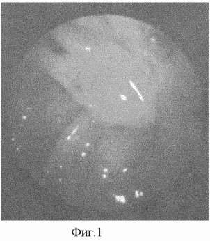
Description of the invention: ultrasound is used as a primary screening procedure in patients with suspected acute cholecystitis. In the ultrasound semiotics of acute obstructive cholecystitis there is a combination of the following main features: an increase in the size of the gall bladder more than 90/30 mm, a thickening of the wall more than 3 mm, the presence of fixed hyperechoic structures with an acoustic shadow in the projection of the cervix of the gallbladder. The second stage of diagnostics is the proposed variant of the CDU. The study is performed 24 hours after the patient enters the hospital. Given that patients with acute cholecystitis after admission are on medical starvation, the study is performed on an empty stomach, and the need for gastric lavage before the study does not arise. Premedication before HDV promedol, atropine sulfate, dimedrol is not carried out. In patients with an increased emotional mood, Seduxen 40 mg is administered intravenously with good effect. The endoscope is administered under local anesthesia with a 10% solution of lidocaine 30 minutes after intravenous injection of 5 ml of a 0.4% solution of indigo carmine. The state of the stomach, duodenum, BSDK, the presence of indirect signs of pancreatitis and cholecystitis are assessed. In the duodenum, 40 ml of a 25% solution of magnesium sulphate is introduced. Further, the arrival of colored bile from the BSDK is expected (FIG. 1). The arrival of turbid bile before the appearance of a colored portion is an objective criterion for the presence of cholangitis. |
When carrying out HDS, there were 4 variants of results in patients with acute cholecystitis:
1) absence of colored bile over 15 minutes after stimulation of cystic reflux by magnesium sulfate;
2) the presence of colored bile in the duodenum without previous stimulation of cystic reflux;
3) appearance of colored bile after stimulation of cystic reflux with magnesium sulfate after 1-6 minutes;
4) the appearance of colored bile from BSDK 6-15 minutes after stimulation.
When comparing the obtained data with operational findings, it was found that during the operations there was an obstruction of the vesicular duct at the first, second and third results of HDS. The most likely cause of early appearance of colored bile is inflammation of the choledocha wall, infection of choledochal bile and, as a result, a change in its pH to the acidic side, which contributes to the oxidation of the leukopathy of indigo carmine and staining of choledochal bile.
Thus, in this situation, the time from the moment of administration of sulfurous magnesia to the appearance of colored bile from the BSDK is important. Clinical and experimental studies have established that the gall bladder normally enters the duodenum after stimulation of bubble reflux with magnesium sulfate after 6-15 minutes. Painted bile, delivered up to 6 minutes, is choleadocic.
In the case of preservation of the bladder duct (confirmed by ultrasound and CDU data), despite the conservative therapy performed within 24 hours, an operation is performed. A method of cholecystostomy using a tube with an obturator balloon is proposed, which is a modification of classical surgical cholecystostomy.
The operation is performed under local anesthesia with neuroleptanalgesia from a small incision in the right hypochondrium. The location of the incision is specified by ultrasound. After the puncture of the bladder, in the region of the bottom, its lumen is opened with a cut to 1.5-2 cm, through which bile and stones are removed. For the drainage of the bladder a rubber tube with a balloon-obturator from latex on the end is used. The reaction of surrounding tissues to the rubber tube is more pronounced than that of polyvinylchloride, which accelerates the formation of the fistulous course.
 |
 |
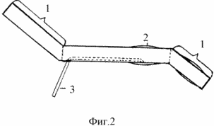
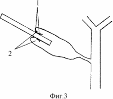
For the production of drainage, rubber intubation tubes of domestic production are used - Fig. 2, where 1 - removed parts of the tube, 2 - balloon obturator, 3 - conductor to fill the balloon. When fixing such a tube, it is possible to confine oneself to applying a single suture seam, which excludes the formation of a "dead space" - FIG. 3, where 1 - the sutures, 2 - "dead space" in the cavity of the gallbladder.
The liquid filled balloon prevents the early prolapse of the cholecystostomy tube and, tightly pressing the bottom of the bladder to the abdominal wall, provides sufficient hermetic activity. To fill the balloon, 0.9% NaCl solution is used. This allows in the future, knowing the initial amount of fluid, in order to prevent pressure ulcers of the gallbladder wall, periodically empty the balloon.
The operation is completed by bringing the safety drainage into the subhepatic space through a separate puncture in the right side region of the abdomen.
In the course of the operation, several factors are necessarily taken into account:
- in order to ensure hermetism, the gallbladder is sutured to the parietal peritoneum;
- if this can not be done, cholecystostoma is formed throughout, with the necessary fixation of the strand of the large epiploon enveloping the tube, to the parietal peritoneum and to the gall bladder. In this case, a tube with a filled balloon becomes particularly important, being a framework for the formation of a fistulous motion;
- The tube inserted into the gallbladder should have a wide lumen ( ![]() 1 cm) for the possible performance of therapeutic and diagnostic endoscopic cholecystoscopy (control of the dynamics of the inflammatory process in the gallbladder, instrumental removal of residual calculi, electrocoagulation of the mouth of the bladder duct).
1 cm) for the possible performance of therapeutic and diagnostic endoscopic cholecystoscopy (control of the dynamics of the inflammatory process in the gallbladder, instrumental removal of residual calculi, electrocoagulation of the mouth of the bladder duct).
In some cases, the development of holostystolism is possible, which is most often associated with overly active behavior of the patient. The presence of safety drainage in the subhepatic space does not always allow to diagnose leakage of bile in time. This is due to the limited drainage operation time (~ 24 hours).
Clinically, it is difficult to suspect a bile duct, The irritant agent is in the immediate vicinity of the operating wound. The use in the postoperative period of analgesics, including narcotic, smooths out peritoneal phenomena. Ultrasonography in the early stages of yellowing and little informative.
In order to control the consistency of cholecystostomy, an original method is used, based on the three-component theory of color vision [42], which assumes that one or another given color is given by three-zone color separation, i.e. Separation of radiation reflected by the object on the blue, green, red ranges of the visible spectrum. With the subtractive principle of color synthesis, the color reproduction is performed by subtraction (substraction) from the white color of the primary colors. The latter is usually achieved by mixing different amounts of colorants on a white or transparent basis, the colors of which are complementary to the main colors - yellow, magenta and cyan, respectively. So the mixture of purple and blue dyes get a blue color (purple color from white subtracts green, and blue - red). Mixing the yellow and magenta dyes gives a red color, and the blue and yellow colors are green (by subtracting the red and blue bands from the white, respectively). Thus, when the additional colors of blue and yellow are blended, one of the primary colors of the visible spectrum is green.
Having always one of the components of this composition - yellow (bile), as the second we have chosen methylene blue. Diluted aqueous solutions of the latter have a blue color. The drug refers to antiseptic products from the group of organic thiosine dyes, non-toxic, does not have a local aggressive effect. A fairly dilute solution is used (up to a clear-blue color). A concentration of 0.05-0.025% is achieved by diluting a standard 1% aqueous solution of methylene blue with physiological saline under visual control.
If there is a suspected incompetence of cholecystostomy, a yellowing in the abdominal cavity, 0.05-0.025% solution of methylene blue is introduced into the safety drainage. The drainage is closed for 5-10 minutes. If the solution stains green (the presence of bile), then, depending on the situation, additional measures are used.
If there is no problem with the consistency of cholecystostomy, within 12-14 days, until the inflammation subsides, the gallbladder is sanitized with antiseptics. With abundant bile cystitis due to cholecystostomy due to bile hypertension due to spasm, inflammatory changes in the sphincter of Oddi, spasmolytics, cholinolytics intramuscularly, warm 25% solution of magnesium sulfate or 0.5% solution of novocaine inward by 1 dessert spoon 10 times a day are used.
Fistuloholegraphy is performed on days 12-14. If the intra- and extrahepatic ducts are not contrasted, provided there are no stones in the gallbladder, its chemical mucoclase is performed. In the case where the cystic duct block is caused by a residual stone, its non-operative removal by chemical dissolution with heparin, alcohol-ether mixture or with the help of various instruments is performed. Manipulation is performed no earlier than 3-4 weeks after the operation. Этот срок необходим для формирования прочного фистульного хода, так как может возникнуть необходимость в манипулировании через свищевой ход.
В случае, когда при фистулографии контрастное вещество поступает во внутри- и внепеченочные протоки, для предупреждения попадания фенола в них, выполняется электрокоагуляция (сила тока 50 мA с экспозицией 10 с) слизистой оболочки устья пузырного протока и прилегающей к нему части шейки пузыря одно- или биполярным электродом под контролем эндоскопа. При наличии стабильного блока, подтвержденного контрольной фистулографией, выполняется мукоклазия.
После выполнения электрокоагуляции возможно возобновление желчеистечения, что обусловлено желчной гипертензией, связанной с нарушением проходимости терминального отдела холедоха (склерозирующий папиллит, аденоматоз БСДК, конкременты и др. ). В таких случаях показана эндоскопическая ретроградная панкреатохолангиография с эндоскопической папиллосфинктеротомией.
Химическая мукоклазия желчного пузыря осуществляется путем введения по трубке 70% эмульсии фенола с экспозицией 4 минуты. Установлено, что именно данная концентрация фенола позволяет добиться полной и безопасной мукоклазии. Применение как менее, так и более концентрированных препаратов фенола, с другой экспозицией приводит к частичной мукоклазии (60%) или некрозу всей стенки (95%). Эффективность воздействия определяется по оценке макроскопических изменений в желчном пузыре и при исследовании микропрепаратов, окрашенных гематоксилином и эозином и по Ван-Гизону.
 |
После аспирации фенола полость пузыря многократно промывается 0,9% раствором хлорида натрия. С целью защиты от химического ожога, кожа вокруг холецистостомы покрывается пленкообразующим клеем или обклеивается лейкопластырем. В дальнейшем в течение 2-3-х недель проводится санация полости желчного пузыря с введением в просвет пузыря мази "Левомеколь", ускоряющей развитие грануляционной ткани. Процесс облитерации контролируется эндоскопическим, сонографическим и рентгенологическим исследованием. Предлагаемый комплексный способ диагностики и хирургического лечения острого холецистита у больных с высоким операционно-анестезиологическим риском, см. предлагаемую схему, был применен у 19 пациентов. Осложнений не наблюдалось. Больные подвергались плановому обследованию через 6, 12 месяцев после выписки из стационара. У всех результаты лечения расценены как хорошие: больные не испытывали болей и неприятных ощущений в правом подреберье и в подложечной области, по данным УЗИ просвет желчного пузыря не определялся, в его проекции имеется плотный эхопозитивный тяж - фиг.4, где А - облитерированный желчный пузырь. |
Преимуществами предлагаемого комплексного способа диагностики и хирургического лечения являются:
- сочетание ХДС и УЗИ в диагностике блока пузырного протока позволяет достичь достоверности, равной 100%, своевременно выставить показания к хирургическому лечению;
- учет временных критериев появления окрашенной желчи при выполнении ХДС позволяет избежать ложноположительных результатов;
- отказ от премедикации промедолом, атропина сульфатом, димедролом исключает артефакты при проведении ХДС;
- применение резиновой трубки с наполняемым баллоном-обтуратором способствует формированию прочного свищевого хода, обеспечивает герметизм холецистостомы, предотвращает преждевременное выпадение дренажа, образование "мертвого пространства" в области дна желчного пузыря и связанные с этим осложнения;
- применение именно 70% эмульсии фенола с экспозицией 4 мин обеспечивает наиболее полную и безопасную мукоклазию желчного пузыря, с последующей полной облитерацией его просвета;
- при выполнении операции исключается отрицательное воздействие на организм пневмоперитонеума и наркоза;
- простота и доступность используемых материалов и самого способа позволяет широко применять его в хирургических стационарах общего профиля.
INFORMATION SOURCES
1. Галеев М.А., Тимербулатов В.М. Желчнокаменная болезнь и холецистит // Уфа,- 1997.-201с.
2. Королев Б.А., Пиковский Д.Л. Экстренная хирургия желчных путей // М., Медицина.- 1990.- 240с.
3. Kukosh MV Clinical course of acute cholecystitis in elderly and senile patients with concomitant diseases // In: Acute cholecystitis. Ways to improve diagnosis and surgical treatment. Sat. Sci. Tr., Gorky. - 1988.- С.50-52.
4. Kochnev OS Emergency surgery of the gastrointestinal tract // Publishing house of the Kazan University. - 1984.-288s.
5. Padishina L. G., Nabegaev A.I., Morozov I.S. Acute cholecystitis in elderly and senile patients // New technologies in surgical hepatology: Mat. 3 conf. Surgeons-hepatol .- St. Petersburg, - 1995.-P.453-456.
6. Boger MM, Mordvov S.A. Ultrasonic diagnostics in gastroenterology // Novosibirsk. Publishing house "Science". Siberian Branch. - 1988.-160s.
7. Grishin I.N. Cholecystectomy: Practice. Allowance // Mn., Vyssh. 1989.-198S.
8. Febres D. - quoted. By L.V. Avdey. Clinic and surgical treatment of cholecystitis // Mn-1963.- 222p.
9. Dederer M.Yu., Krylova NP, Ustinov G.G. Gallstone disease, M., Meditsina.-1983.-176c.
10. Dikhtenko G.I. Accelerated chromatic duodenal sounding in express diagnostics of acute cholecystitis // Klin. Chir.- 1971.- 3.-C.13-16.
11. VV Vakhidov, M. Khodzhibekov, K. Tsoi. Echography and cholecystography in the diagnosis of calculous cholecystitis // Vest.khir.- 1984.- 11.-P.35-38.
12. Postolov PM, Bykov AV Ultrasonic semiotics and diagnostics of acute cholecystitis // Surgery. -1990.- 2.-P.21-23.
13. Kochnev OS, Kim IA, Valeev AG Endoscopic diagnosis and treatment of acute cholecystitis // Surgery.-1984. - 7.- P.25-30.
14. Mashkovskiy M.D. Medicines: in 2 volumes.-10th ed. Sr. -M.-1985.-624s-T.2.-576s.
15. Zatevakhin II, Kushnir VK, Tsitsiashvili M.Sh., Blinov V.Yu., Ugolnikov S. G. Endoscopic cholecystostomy in the treatment of acute cholecystitis in persons with a high degree of operational risk // Surgery.- 1988 .- 1.-C.11b-117.
16. AP Chadaev, AS Lyubsky. Two-stage treatment of acute cholecystitis in elderly patients // Intern. Sci. Conf .: "Actual problems of diagnostics and treatment of diseases of the hepatobiliary zone." Endoscopic surgery. " Dokl.-SPb. 1996.-pp. 162-163.
17. Braun, B., Blank, W. Gallbladder, puncture and drainage as therapy of cholecystitis, Med. Klin. 1996. Vol. 91, 6.-P.359-365.
18. Briskin BS, Minasyan AM, Vasilyeva MA, Barsukov MG. Percutaneous transhepatic microcholecystostomy in the treatment of acute cholecystitis // Annals of surgical hepatology .- 1996.- 1.- P.98-107.
19. Verbanck JJ, Demol JW, Ghillebert GL et al. Ultrasound quide puncture of the gallbladder for aqute cholecystitis // Lancet.- 1993.- 341.- P.1132.
20. Van Steenbergen W., Ponette E., Marchal G. et al. Percutaneous transhepatic cholecystostomy for acute complicated cholecystitis in elderly patients // Am. J. Gastroenterol .- 1990.- 85.- P. 1363.
21. Van Overhagen H., Meyers H., Tilanus HW, Jeekel J., Lamberis JS Percutaneous cholecystostomy for patient with acute cholecystitis and an increased surgical risk. // Cardiovasc. Intervent. Radiol. 1996.-Vol. 19, 2.-P.72-76.
22. England RE, McDermott VG, Smith TP, Suhocki PV, Payne CS, Newman GE "Percutaneous cholecystostomy: who responds?" // Am. J. Roentgenol .- 1997.- Vol.168, 5.- P. 1247-1251 .
23. Bakke K., Navjord D., Nilsen BH Percutaneous drainage of the gallbladder in acute cholecystitis // Tidsskr / Laegeforen .- 1999, -Vol. 119, 22.-P. 3260-3262.
24. Ohotnikov OI Percutaneous transhepatic microcholecystostomy under echoscopic control in complex therapy of acute cholecystitis in patients with high operational risk // Morphogenesis and Regeneration: Mat. Conf. Morphol. , Immunol. And Chernozemye clinicians, pos. 60th Anniversary Course. State. honey. In-ta .- Kursk .- 1995.- P.62-63.
25. Shapoval'yanets SP, Mikhayusov SV, Maksimova VV Indications for microcholecystostomy under ultrasound control // Surgery.-1997.- 1, -C.68-71.
26. Famulari S., Macri A., Galipo S., Terranova M., Freni O., Guzzocrea D. The role of ultrasonographic percutaneous cholecystostomy in treatment of acute cholecystitis // Hepatogastroenterol .- 1996.- Vol.43, 9. - P. 538-541.
27. Sugiyama M., Tokuhara M., Atomi Y. Is percutaneous cholecystitis in the very elderly? / / World J. Surg.-1998.-Vol. 22, 5.- P. 459-463.
28. Dumas R., Caroli-Bosc FX, Demarquay JF Zanaldi H., Hastier P., Conio M., Maes B., Delmont JP Acute inoperable cholecystitis treated by endoscopic naso- vesicular drainage. Study of 15 patients // Gastroenterol / Clin. Biol.-1997.-VOL.21, 11.-P. 854-858.
29. Hu-hai, Xue-shou, Wang Xue-zhi, Zhang Sheng-dao. Extracorporeal shokwave lithotripsy and methyl tert-butyl ether for gallbladder stones // Chin. Med. J.- 1992.-Vol.105, 8.- P. 630-634.
30. Barton KE, Picus D., Hicks ME, Darcy MD, Vesely TM, Kleinhoffen MA Electrohydraulic lithotripcy as an a djunct to percutaneous and endoscopic removal of biliary calculi // 17 th Annu. Sci. Meet., Washington. DC : Program / Soc. Cardiovasc. and Intervent. Radiot- Washington (DC). -1992.-P. 47.
31. Королев Б. А., Пиковский Д.Л., Грудинская И.Н. Холецистостомия при остром холецистите // М., Медицина.- 1973.-104 с.
32. Дзарасова Г.Ж. Отдаленные результаты двухэтапного эндоскопического метода лечения острого калькулезного холецистита у больных с высокой степенью операционного риска // Автореф. дис....канд. honey. Sciences. М.,-1993.
33. Емельянов С. И., Федоров А.В., Феденко В.В., Матвеев Н.Д., Евдокименко В.В., Александров К.Р. Технологические аспекты эндоскопической хирургии желчных путей // Анналы хирургической гепатологии.-1996.- 1.-С.115-119.
34. Гуляев А.А., Шаповальянц С.Г., Бурова В.А., Михайлусов С.В., Аввакумов А.Г. Облитерация просвета желчного пузыря у. больных с высоким операционным риском // Хирургия.-1998.- 9.- С.42-44.
35. Ji ZL, Chen HR, Lei RQ, Yang JZ, Huang MH Cystic duct occlusion by microwave tissue coagulator in rabbits // J/R Coil. Surg. Edinb.- 1991.-Vol. 36, 6.-P. 395-398.
36. Никуленков С.Ю., Бельков А.В., Ефимкин А.С. Эндоскопическая облитерация желчного пузыря у больных острым холециститом с высоким операционным риском // Эндоскопическая хирургия.-1998.- 1.-С.34.
37. Шуркалин Б.К., Ермолов А.С., Гуляев А.А., Удовский Е.Е., Юрченко С. В. , Каримов TM, Пономарев В.Г., Елисеенко В.И., Лурье Б.Л. Химическая мукоклазия и облитерация просвета желчного пузыря у больных с высоким операционным риском (экспериментальное исследование) // Хирургия.- 1993.- 4.-С. 38-43.
38. Назаренко П.М., Тарасов О.Н., Должиков А.А. К морфологическому обоснованию метода облитерации желчного пузыря при лечении желчнокаменной болезни // Морфогенез и регенерация: Мат. Conf. морфол., иммунол. и клиницистов Черноземья, посв. 60-летию Курс. State. honey. ин-та.-Курск.- 1995.-С. 59-61.
39. Becker CD, Fache JS, Malone DE, Stoller JL, Burhenne HJ Ablation of the cystic duct and gallbladder: clinical observations // Radiology.- 1990.-Vol. 176, 3.-P. 687-690.
40. Girard MJ, Saini S., Mueller PR, Lee MJ, Ribeiro RE, Fermcci JT, Flotte TJ Percutaneous chemical gallbladder sclerosis after laser - induced cystic duct obliteration: results in an experimental model // Am. J. Roentgenol.-1992.- Vol.159, 5.- P. 997-999.
41. Girard MJ, Saini S., Mueller PR, Flotte TJ, Staritz M., Domankevitz Y. , Ferrucci JT , Nishioka N. Percutaneous obliteration of the cystic duct with a holmium: yttrium - aluminium - garnet laser: results of in vitro and animal experiments // Am. J. Roentgenol.- 1992.- Vol.159, 5.- P. 991-995.
42. Б.С.Э. Издательство: Советская энциклопедия.-1978.- т. 28.- С. 460.
CLAIM
Комплексный способ диагностики и хирургического лечения острого холецистита у больных с высоким операционно-анестезиологическим риском, включающий ультразвуковое исследование, хромодуоденоскопию, хирургическую холецистостомию, химическую мукоклазию, отличающийся отсутствием премедикации при выполнении хромодуоденоскопии у больных с нормальным эмоциональным настроем и использованием внутривенно 40 мг седуксена у больных с повышенным эмоциональным настроем, применением магния сульфата для стимуляции пузырного рефлюкса, учетом временного фактора выделения окрашенной желчи из фатерова соска в двенадцатиперстную кишку при диагностике проходимости протока, с нормой ее поступления через 6-15 мин после стимуляции, использованием холецистостомии под местной анестезией в случае сохранения блока пузырного протока при помощи трубки с просветом более или равным 1 см, с баллоном-обтуратором, фиксируемой одним кисетным швом, исключающей отрицательное воздействие на организм наркоза, пневмоперитонеума и образование "мертвого пространства" в зоне кисетных швов, заполнением баллона жидкостью, введением в страховочный дренаж 0,05- 0,025% раствора метиленовой сини для ранней диагностики и контроля состоятельности холецистостомы, и при получении из дренажа отделяемого зеленого цвета, проведении мероприятий, устраняющих несостоятельность холецистостомы, проведением химической мукоклазии 70% эмульсией фенола, с экспозицией 4 мин, сохранением холецистостомической трубки до окончания отхождения некротизированной слизистой оболочки, использованием для уменьшения желчеистечения из холецистостомы медикаментозных препаратов, направленных на уменьшение отека и спазма фатерова соска.
print version
Date of publication 28.01.2007gg


Comments
When commenting on, remember that the content and tone of your message can hurt the feelings of real people, show respect and tolerance to your interlocutors even if you do not share their opinion, your behavior in the conditions of freedom of expression and anonymity provided by the Internet, changes Not only virtual, but also the real world. All comments are hidden from the index, spam is controlled.