
| Start of section
Production, amateur Radio amateurs Aircraft model, rocket-model Useful, entertaining |
Stealth Master
Electronics Physics Technologies Inventions |
Secrets of the cosmos
Secrets of the Earth Secrets of the Ocean Tricks Map of section |
  |
| Use of the site materials is allowed subject to the link (for websites - hyperlinks) | |||
Navigation: => |
Home / Patent catalog / Catalog section / Back / |
|
INVENTION
Patent of the Russian Federation RU2036235
![]()
METHOD OF INACTIVATING VIRUSES IN BLOOD AND ITS COMPONENTS
The name of the inventor: Harald Mor [DE]; Bernd Lambrecht [DE]
The name of the patent holder: Bluthspindeinster der Landesferbende Des Dochenchen Rothen Croits Niedersachsen, Oldenburg und Bremen GBM (DE)
Address for correspondence:
The effective date of the patent: 1992.03.1
Use: virology, biotechnology.
SUMMARY OF THE INVENTION: Inactivation of viruses in the blood and its components is carried out by mixing the treated solution or suspension with phenothiazine dye and subsequent irradiation with light, wherein the phenothiazine dye is used in a concentration of 0.1 to 10 μmol, the irradiation being carried out directly in clear containers used for collection And storage of blood.
DESCRIPTION OF THE INVENTION
The invention relates to the control of viruses, in particular the inactivation of viruses in the blood and its components.
A method for inactivating viruses is known in which viruses in the form of a liquid are mixed in a flask with a phenothiazine dye followed by light irradiation (see V. Snipe et al., 1979 Photochem., And Photobiol. 29, pp. 785-790). When a known method was used to inactivate viruses in the blood and blood components, it was found that in this case, inactivation not only of viruses but also of blood plasma proteins, such as coagulation factors, occurred.
The purpose of the invention is to develop a simple method of inactivating viruses in the blood and its components, in which various kinds of viruses are killed without significant functional influence on blood plasma proteins.
The task is solved in a method for inactivating viruses in the blood and its components by mixing the treated solution or suspension with a phenothiazine dye and subsequent irradiation with light due to the fact that the phenothiazine dye is used in a concentration of 0.1-10 μmol, the irradiation being carried out directly in transparent containers serving For taking and storing blood. The irradiation is carried out with daylight of sufficient intensity or monochromatic light, preferably a source of cold light having a wavelength in the region of absorption maximum of the corresponding dye. In addition, when the viruses are inactivated in the blood plasma or in solutions of plasma proteins, the following conditions must be observed. The operating temperature should be 0-37 ° C, but possibly 4-20 ° C. The inactivation process lasts, in particular, from 5 minutes to 5 hours, preferably from 10 minutes to 3 hours. The pH of the medium to be treated should be 5- 9, preferably 6-8.
It was found that a non-enveloped virus, such as an adenovirus that can not be inactivated in a plasma under physiological conditions, can be photosensitized by freezing and subsequent thawing, after which inactivation can be successfully carried out. In this case, inactivation can be established regardless of the sequence of freezing techniques and the subsequent thawing and addition of the dye. Freezing refers to the process of deep freezing of liquefied gases as a refrigerant at a temperature of about -20 ° C to 80 ° K. As a rule, the freezing is carried out at a temperature of -30 ° C.
Inactivation of viruses can be carried out directly in bags designed to store blood or blood plasma, although these bags have only limited light transmittance. In this case, the proposed method is reduced only to the addition of a dye, followed by irradiation of the bag together with its contents. Then, the corresponding product can be further processed.
Thus, the proposed method can be carried out by simple technical means and therefore it can be integrated into the technological process of processing the donor blood in the respective stores. An insignificant amount of the added dye can remain in the further processed liquid. If necessary, it can be removed using an adsorber. The concept of blood and its components is understood to mean: whole blood, red blood cell concentrates, platelet concentrates, blood plasma, whey, cryo-sediment, coagulation factor concentrates, inhibitors, fibronectin, albumin.
In the method of the present invention, phenothiazines of the following structural formula
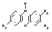
In Table. 1 shows the values of X, R 2 , R 3 , R 7 .
Example 1
Photoinactivation is subjected to the vesicular stomatitis virus in the human blood plasma in the presence of methylene blue. About 5 × 10 7 spot-forming units per ml of the virus are suspended in human blood plasma and mixed with methylene blue, used in various concentrations. Control samples do not contain dye. The sample volume is 0.5 ml. One control sample and part of the methylene blue containing samples are irradiated with visible light at room temperature for 4 hours. The remaining samples are stored for the same period of time. The light source is a diaprojector equipped with a 150 W halogen lamp. The distance between the lens of the diaprojector, i.e., the light output port, and the samples in all the experiments is 30 cm (excluding the case of inactivation of viruses in blood bags). After irradiation, a virus titer is determined in all samples by spot analysis. As an indicator, BHK cells (ATCC N CCSS10) were dried. The results of the experiments are summarized in Table. 2.
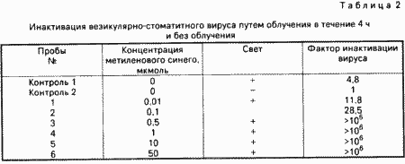
The data of Table. 2 indicate that, starting with a concentration of methylene blue equal to 0.5 μmol, the infectious titer of the virus decreases by more than 6 decimal degrees.
Example 2
This experiment confirms inactivation of the virus at low dye concentrations.
Aliquots of blood plasma with a volume of 500 μl containing the vesicular stomatitis virus and various amounts of methylene blue are irradiated by means of a 30 cm wide projector in the refrigerating chamber. Samples A-E are irradiated, and the G-sample remains unirradiated.
The results of the experiment are summarized in Table. 3. They show that under the given conditions the specified virus is inactivated by more than 4 decimal degrees. This requires a concentration of methylene blue, equal to 0.5 μmol. One overnight incubation at 4 ° C probably leads to a decrease in the virus titer by 1-2 decimal degrees, which may be an explanation for the relatively low initial titer. But this circumstance has not been studied in detail.
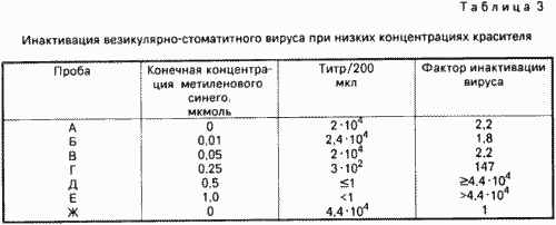
Comparison of the results of inactivation of irradiated sample A with sample G (only storage in the dark) shows that one light obviously does not have a big effect on the infectivity of the virus.
Example 3
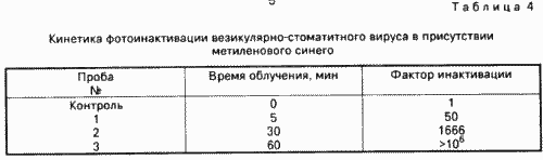
The dependence of photoinactivation of viruses in the presence of phenothiazine dyes on the duration of irradiation is studied. In this case, 10 6 spot-forming units per ml are suspended in blood plasma and the samples are irradiated in the manner described in the previous examples at 22 ° C for the time indicated in Table 1. 4 times. According to Table. 4 that under these conditions one-hour irradiation is sufficient to reduce the infectious titer of the vesicular-stomatitis virus by more than 6 decimal degrees.
Example 4
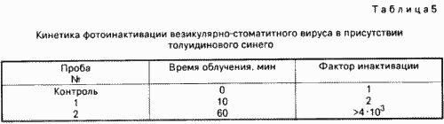
Example 3 is repeated with the difference that the inactivation is carried out in the presence of 1 μmol toluidine blue. The results of the experiment, summarized in Table. 5, suggest that the vesicular stomatitis virus can effectively be inactivated in the presence of toluidine blue.
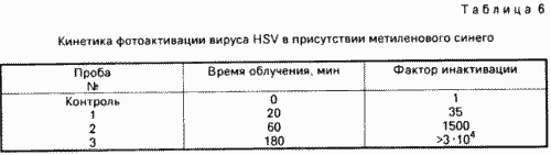
In the presence of phenothiazine dyes, it is possible to inactivate the HSV vesicle virus, as well as the HIV-1 AIDS virus, as evidenced by the results of Examples 5 and 6.
Example 5
The inactivation of the HSV virus is carried out in the presence of 1 μmol of methylene blue. In Table. 6 data on the kinetics of photoinactivation of HSV virus are summarized.
Example 6
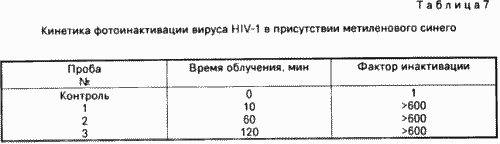
Inactivation of the HIV-1 virus is carried out in the presence of 1 μm methylene blue. The virus titer is 6 × 10 2 spot-forming units / ml and MT4 cells (HTLV-1 infected human T-lymphoblastoid cell line) are used as an indicator. The results of the experiment, summarized in Table. 7, indicate that the HIV-1 virus is very sensitive to photoinactivation, since already during the first 10 minutes the virus titer decreases more than 600 times.
Example 7
Inactivation of non-enveloped viruses under ordinary physiological conditions in the presence of 80% plasma was unsuccessful. In this case, an adenovirus was used as an example of a virus without a cladding, which is preincubated for the period of time indicated in the table. 8 times at 4 ° C in the dark in the presence of 1 μmol methylene blue. Then, a 30-minute irradiation with a halogen lamp of 150,000 lux is carried out. The infectivity of adenovirus remains unchanged. The results of the experiment are summarized in Table. 8.
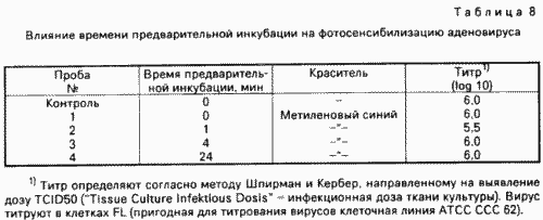
When toluidine blue is used under the same experimental conditions, there is no decrease in the virus titer.
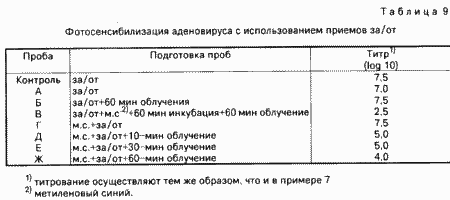
To achieve inactivation of the adenovirus, the processes include freezing and thawing techniques (hereinafter: "for / from"), while freezing is carried out to -30 ° C. The sequence of steps for / from and the moment of addition of the dye (1 μmol methylene blue) play only a secondary role . Irradiation of samples is carried out with a halogen lamp of 120,000 lux. The results of the experiment are summarized in Table. 9.
Example 8
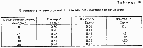
This experiment is aimed at studying the effect of dye in various concentrations on the activity of clotting factors. At the same time, 2 ml aliquots of human blood plasma are mixed with various amounts of methylene blue. Immediately after the addition of the dye, the activity of coagulation factors V, VIII and IX is measured. As can be seen from the data in Table. 10, all clotting factors are inhibited depending on the concentration of the dye, i.e. the inflection of factors VIII and V begins at a concentration of about 10 μm, and factor IX is already inhibited by the presence of a dye at a concentration of about 2.5 μm. Thus, methylene blue at higher concentrations directly affects proteins without exposure to light.
The results of the experiment are summarized in Table. 10.
Example 9
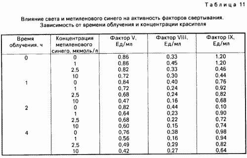
The activity of clotting factors is affected not only by the concentration of the dye, but also by the time of irradiation. This dependence is investigated in this example. To this end, aliquots (2 ml) of human blood plasma are mixed with various amounts of methylene blue and irradiated in the manner described in Example 1 for 1-4 hours. Control samples are not subjected to photographic processing. The data of Table. 11 show that the activity of the three clotting factors V, VIII and IX is inhibited depending on the irradiation time and the dye concentration. In particular, data on coagulation factors VIII and IX indicate that a higher concentration of methylene blue and light irradiation with a duration of 2 h leads, apparently, to an increase in the thrombolytic effect.
Example 10
According to the invention, the photoinactivation of viruses is carried out directly in transparent containers serving for the collection and storage of blood or its components, after preliminary addition of the dye in the required amount. In this simple way, it is possible to process blood and its components of individual donors at any time.
In this example, three samples of fresh human blood plasma are thawed, to which 1.5 × 10 6 stain-forming units of the vesicular stomatitis virus are added in their containers. To two samples, methylene blue is added at a concentration of 1 and 10 μmol, respectively. A sample that does not contain methylene blue is taken as a positive control in the dark at 4 ° C. Then all the containers are clamped between two plexiglass plates in order to provide an even layer thickness of about 2.5 cm. The containers are irradiated with a diaprojector, Placed at a distance of 90 cm. After 4 hours, samples are taken, which for determination of the virus titer are fed to FL cells. The results of the experiment are summarized in Table. 12.
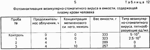
The data of Table. 12 indicate that already a concentration of 1 μmol of methylene blue is sufficient to reduce the infectious titer of the vesicular stomatitis virus by more than three decimal degrees. Even in the absence of a dye, irradiation leads to a decrease in the virus titer, but only by about 50%
At concentrations up to 1 μmol, the phenothiazine dyes used for virus inactivation can remain in the blood or its components without any side effects. But they can also be removed by dialysis, gel filtration or adsorption. The most interesting is adsorption, since it is associated with the least time and technology and, in addition, the corresponding protein solutions do not need dilution. It was found that methylene blue and other phenothiazine dyes bind to various commercially available separation gels, including those that do not have the ability to bind proteins or have only a poorly developed ability to bind them. Thus, such adsorbents are particularly suitable for subsequent removal of the photo-oxidant. Among the adsorbents investigated for separating methylene blue and other phenothiazine dyes, the agents listed in Table 1 can be used successfully. 13.
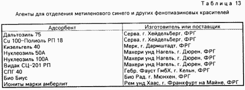
In most cases, 2 g of the corresponding adsorbent is sufficient for complete extraction from the solution of plasma proteins of methylene blue in concentrations of 10 μmol. The most suitable were two types of adsorbents.
1. Silicagels with a pore diameter of 40-100 ![]() , Which do not allow plasma proteins to penetrate into the matrix. But molecules of low molecular weight dyes reliably bind due to ionic, electrostatic and hydrophobic interactions. Examples of such commonly available adsorbents are: Matrex Silica Gel of the foreign company Amicon, Witten, FRG, Daltozol of Servo, Heidelberg, West Germany, and kieselgel of the foreign company Merck, Darmstadt, Germany.
, Which do not allow plasma proteins to penetrate into the matrix. But molecules of low molecular weight dyes reliably bind due to ionic, electrostatic and hydrophobic interactions. Examples of such commonly available adsorbents are: Matrex Silica Gel of the foreign company Amicon, Witten, FRG, Daltozol of Servo, Heidelberg, West Germany, and kieselgel of the foreign company Merck, Darmstadt, Germany.
2. Gels based on polystyrene and divinylbenzene or polymers of esters of acrylic acid. Examples of commercially available gels of these types are amberlites produced, for example, by the foreign company Rem Haund Haas, Frankfurt am Main, Germany, and the bio bidus of the Bio Rad company in Munich, Germany. They mainly serve to remove non-polar or surfactants, for example detergents, from aqueous solutions. They are nonpolar or only weakly polar.
Example 11
Fresh blood plasma is mixed with 10 μmol methylene blue (mc). Aliquots of 5 ml volume are mixed with various amounts of daltozil (pore diameter 75 ![]() ) And bio bidus SM 16 (pore diameter 144
) And bio bidus SM 16 (pore diameter 144 ![]() ) Respectively, followed by stirring for 30 minutes. Then the gel is allowed to settle. In the blood plasma, the content of factor VIII and factor V is determined, extinction at 660 nm, and the protein content of some samples.
) Respectively, followed by stirring for 30 minutes. Then the gel is allowed to settle. In the blood plasma, the content of factor VIII and factor V is determined, extinction at 660 nm, and the protein content of some samples.
The results of the experiment are summarized in Table. 14.
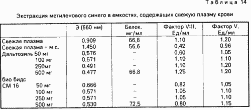
According to the extinction data, it is evident that along with the colorant from the plasma, other substances are obviously extracted, which, however, are not plasma proteins. Data on the extinction of plasma treated with 100-250 mg of adsorbent per 5 ml, i.e. 2-5 w / v volume, hardly differ from those extinction data of the samples, which were extracted with 10 wt. Adsorbent / sample volume. Therefore, when the concentration of methylene blue is 10 μm, it is sufficient to use the adsorbent in an amount of 2-5% by weight of the sample so that in the case of a batch process, the colorant is completely removed from the plasma. At lower dye concentrations, a correspondingly lower consumption of the adsorbent is required.
Example 12
In this example, instead of blood plasma, use a 5% solution of human serum albumin (hereinafter: ACH). In this test, the concentration of methylene blue is 10 μmol. Aliquots of 5 ml volume are extracted with 100 mg (2% by weight) of the following adsorbents: daltosyl (pore diameter 75 ![]() ), Silica gel (pore diameter 40
), Silica gel (pore diameter 40 ![]() ) And bio bidus SM 16 (pore diameter 144
) And bio bidus SM 16 (pore diameter 144 ![]() ).
).
The results of the experiment are shown in the drawing. It shows that in all cases the extinction decreases at 660 nm, i.e. This time is sufficient for the periodic removal of the photo-oxidant from the protein solution. It can be seen from the drawing that in this case the adsorbents of the bio-bidus SM 16 and silica gel with a pore diameter of 40 ![]() Are more effective than daltosyl with a pore diameter of 75
Are more effective than daltosyl with a pore diameter of 75 ![]() .
.
Example 13
In this example, the possibility of adsorption removal of methylene blue from a solution of plasma proteins by column chromatography is investigated. This method is based on the idea that after the inactivation of viruses by means of dye and irradiation, the solution of plasma proteins can be passed through a column with an adsorbent and from there to be fed to a further container serving to store the treated solution. In this case, the column can be placed between the two tanks. Thus, it is possible to pre-manufacture a unit consisting of two containers with an adsorption column placed between them. Thus, it is possible to create a closed system, which greatly reduces the risk of contamination of the inactivated virus of a preparation of plasma proteins, including preparations from individual donors.
This experiment is carried out as follows.
250 ml. A 5% solution of albumin is passed at a different rate through a column containing 5 ml of silica gel with a pore diameter of 40 ![]() . Fractions of 10 ml were collected and subjected to extinction at 660 nm. According to Table. 15 it can be seen that the whole albumin solution can be passed at a rate of 5 and 7.5 ml / min, respectively, through the column, without the residual methylene blue being shown in the fractions obtained. Therefore, removal of the dye from a solution of 250 ml requires no more than 30-35 minutes.
. Fractions of 10 ml were collected and subjected to extinction at 660 nm. According to Table. 15 it can be seen that the whole albumin solution can be passed at a rate of 5 and 7.5 ml / min, respectively, through the column, without the residual methylene blue being shown in the fractions obtained. Therefore, removal of the dye from a solution of 250 ml requires no more than 30-35 minutes.
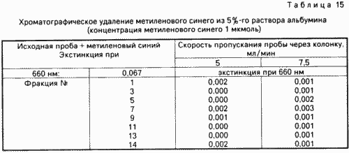
The results of this experiment show that chromatographic removal of the photo-oxidant can be carried out without any problems. On the other hand, the aforementioned possibility of obtaining inactivated virus preparations of plasma proteins from individual donors has been proved.
CLAIM
A method for the inactivation of viruses in the blood and its components by mixing the treated solution or suspension with a phenothiazine dye and subsequent irradiation with light, characterized in that the phenothiazine dye is used at a concentration of 0.1 to 10 μmol, the irradiation being carried out directly in clear containers for collection and storage Blood.
The method according to claim 1, wherein the phenothiazine dye is toluidine blue or methylene blue.
The method according to claim 1 or 2, characterized in that the irradiation is carried out by visible light in the region of absorption maximum of the corresponding dye.
Method according to one of the claims. 1 3, characterized in that the irradiation is carried out at a pH of the medium of 5 9.
The method according to any one of claims 1 to 4, characterized in that the irradiation is carried out for 5,300 minutes.
The method according to any one of claims 1 to 5, characterized in that the irradiation is carried out at 0-37 ° C.
A method according to any one of claims 1 to 6, characterized in that the treated solution or suspension is subjected to a preliminary freezing, followed by thawing before irradiation.
The method of claim 7, wherein the dye is added prior to the freezing process.
The method of claim 7, wherein the dye is added after thawing.
A method according to any one of claims 1 to 9, characterized in that, after irradiation, the blood or components thereof is passed through an adsorbent to remove the colorant.
The method according to claim 10, characterized in that it uses two containers suitable for taking blood, with an adsorption column placed between them.
The process according to claim 10 or 11, characterized in that the adsorbent is an agent from the group consisting of silica gel, polystyrene-based divinylbenzene-based gel, and acrylic acid ester polymers.
print version
Date of publication 08.11.2006gg
![]()

Comments
When commenting on, remember that the content and tone of your message can hurt the feelings of real people, show respect and tolerance to your interlocutors even if you do not share their opinion, your behavior in the conditions of freedom of expression and anonymity provided by the Internet, changes Not only virtual, but also the real world. All comments are hidden from the index, spam is controlled.