|
Start of section
Production, amateur Radio amateurs Aircraft model, rocket-model Useful, entertaining |
Stealth Master
Electronics Physics Technologies Inventions |
Secrets of the cosmos
Secrets of the Earth Secrets of the Ocean Tricks Map of section |
  |
| Use of the site materials is allowed subject to the link (for websites - hyperlinks) | |||
Navigation: => |
Home / Patent catalog / Catalog section / Back / |
|
INVENTION
Patent of the Russian Federation RU2254830
![]()
DEVICE FOR STRETCHING AND / OR ASSEMBLING HOLES OF HEART VALVES (VARIANTS) AND METHOD OF CORRECTION OF SIZES AND / OR CONSOLIDATION
HOLE OF HEART VALVES
The name of the inventor: KALANGOS Afksendiyos (CH); ANDREI Raymond (CH); Le Goff Philippe (CH)
The name of the patent owner: BIORING S.A. (CH); CALANGOS Afxendiyos (CH)
Address for correspondence: 129010, Moscow, ul. B. Spasskaya, 25, p. 3, LLC "Gorodissky & Partners Law Firm", Yu.D. Kuznetsov
Date of commencement of the patent: 2000.11.07
The invention relates to medicine and can be used in cardiosurgery to correct the size and / or enhancement of the heart valve openings. The device comprises a connecting member of a resorbable material and is connected at one of its ends to a thread which provides a longitudinal pulling force for inserting the connecting member into the fabric of the valve ring for mounting. The second version of the device is characterized by the needle. The needle is inserted into the endomyocardium of the valve rings, passed through a part of the perimeter of the hole, the needle is withdrawn, the thread is guided through the endomiocardium with the help of a thread so that the free end of the connecting element is at the point of injection, fixed in the endomyocardium, the connecting element is passed through the ring to that position , When the other end will be located at the extraction point and exit the endomyocardium. Fix the second end. The invention allows to ensure the normal growth of the valve ring in children.
DESCRIPTION OF THE INVENTION
The present invention relates to a device for contracting and / or reinforcing the openings of valvular valves and a method for correcting the dimensions of the openings of the valvular valves.
Defects in the valve openings of the heart, whether sigmoid valves of the aorta or pulmonary artery, or mitral valves, or tricuspid valves, in about 80-90% of cases are the result of prolapse (prolapse) or narrowing of the valves, which entails widening of the valve ring, The volumes of the corresponding heart cavities, and in almost all other cases is a consequence of the expansion of this valve ring without damaging the associated valve.
After correcting the defects of the corresponding valves, it is necessary to simultaneously correct the expansion of the ring containing this valve and to keep the ring in its normal dimensions. To prevent possible recurrence of such defects, it is necessary to reinforce the ring surrounding the valve openings.
To correct such apertures containing valves, many different types of rigid or flexible ring-shaped implants have been proposed, for example, as DURAN, CARPENTIER or PUIG-MASSANA rings, or segments such as COSGROVE. These rings or segments rigid or flexible are placed and fixed by sewing along the perimeter of the ring to be corrected and containing the valve. Thus, the valve-containing orifice is brought to the desired size, usually calculated in an appropriate proportion of the patient's body surface area, and maintained at this normal size.
EP 0338994 describes an apparatus for surgically repairing a tricuspid heart valve deficiency used for fixation by specific stitching along the perimeter of the ring of the valve-containing orifice to be corrected, which occurs on the inner surface of this opening. To speed up the process of fixation by applying a seam, the device can be equipped at the ends with a thread and a needle. However, the thread and the needle, respectively, serve no other purpose than that determined by the desire to provide faster cross-linking after completion of the insertion of the device into the heart, and the device remains within the already known implants and implantation technologies, and only the speed of performing conventional surgical operations is improved.
Rings for implantation are usually made of synthetic material or metal and in some cases lead patients to valve infections in case of bacteremia requiring preventive therapeutic treatment and, if necessary, new surgical intervention.
In addition, when implantable rings in the body are rigid or flexible, they are used to operate children or infants, they prevent the normal growth of the corresponding ring of the heart opening containing the valve, leading to stenosis, as well as to one or more consecutive new surgical interventions , Necessary for the expansion of this ring and for the replacement of a stenotic heart valve.
Patent publication WO 97/16135 describes an annular heart prosthesis that can be resorbed in a living body. The annular prosthesis is made of biodegradable material in order to allow the material to be replaced by the patient's own biological material during the dissolving of this annular prosthesis. However, the ring prosthesis is designed to be placed in the usual manner on the inner surface of the valve-containing orifice along the perimeter of the ring of this correction hole.
It is an object of the present invention to provide an apparatus for contracting and / or reinforcing heart-containing valves that will exclude any predisposition to infection and ensure a normal growth of the valve ring in children, thereby avoiding stenosis or sequential surgical interventions. This device is designed in such a way as to provide the possibility of a completely new and innovative surgical movement and greatly improve the speed, simplicity and applicability of surgical intervention.
The device for contracting and / or reinforcing the heart holes containing the valves is characterized by the features indicated in claim 1 and / or in claim 2.
In the following, the invention is illustrated by a description of preferred embodiments of the device with reference to the accompanying drawings in which:
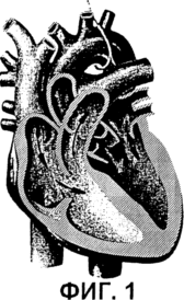
1 shows a general view of the human heart
 |
 |
 |
 |
 |
 |
 |
 |
|
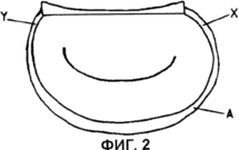
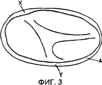
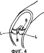
Figures 2, 3 and 4 show simplified, respectively, mitral valve, tricuspid valve and sigmoid heart valves;
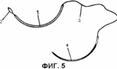
5 shows schematically a first embodiment of a constriction or reinforcement device according to the invention;
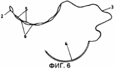
FIG. 6 is a schematic view of a second embodiment of a constriction or reinforcement device according to the invention; FIG.
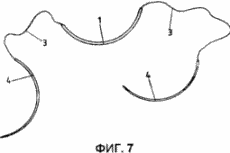
FIG. 7 shows schematically a third embodiment of a constriction or reinforcement device according to the invention; FIG.
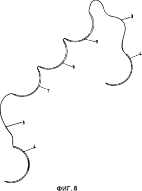
FIG. 8 shows schematically a fourth embodiment of a constriction or reinforcement device according to the invention;
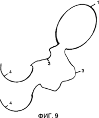
FIG. 9 is a schematic representation of an embodiment of the apparatus of FIG. 7 according to the invention. FIG.
Instead of rigidly and / or finally fixing the ring or segment rigidly or flexibly along all or part of the perimeter of the hole containing the valve, as it is done up to now, it is proposed to place a flexible connecting element along all or part of the perimeter of the valve-containing orifice within the endocardium or layer Tissue located on the inside of the muscle of the myocardium, determine the size of the opening of the valve opening with a certain length of this connecting element and fix the connecting element at one or two points of the endocardium, for example, using sutures.
In addition, according to the invention, a resorbable body element (or element that is biodegradable or biologically absorbable), i.e., biodegradable and not causing an immune response from the body, is used. In the following, the term "resorbable material" is used to define a biologically absorbable or biodegradable material and the term "sutures" that can be resorbable or not resorbable in a living body. At the first time after the operation, the connecting element maintains the valve-containing ring at normal or desired sizes, preventing the expansion of this ring.
Then, the material of this connecting element dissolves within the endomyocardial layer, the organism creates a scar in the form of reaction along this connecting element, characterized by a fibrous tissue representing a higher tensile strength. Thus, after the connecting member is absorbed by the body, the retention of the valve opening at the desired size is achieved by the rigidity of the fibrous scar tissue.
Since the residual scar is formed by the patient's own biological tissues, there can not be a predisposition to infection, but it is especially important that this scar can grow normally during the growth of the child, which eliminates the problems of late stenosis.
The new technology is possible thanks to the device of tightening and / or reinforcement for the correction of defects. In a first embodiment, the device comprises a connecting member 1 made of a material that is resorbable in a living body, terminating at one end of the loop 2 or by a locking and locking organ that is either resilient or resilient, such as a fishing hook, This end to the endomyocardium. The other end of the connecting element 1 is fastened, usually during the manufacture of the device, with a thin and very flexible thread 3 of the type of thread to perform surgical sutures. The fine thread 3 is resilient and is usually made of the same material as the connecting element 1.
The thread 3 is attached by its free end to the needle 4 and provides the possibility of mounting on the intended place of the connecting element 1 of this device.
Obviously, the proposed device is implemented in numerous sizes, since for convenient installation it is preferable that the curvature and length of the needle 4 correspond to the curvature of the ring of the hole containing the valve and the length of the perimeter portion of this hole that must be provided with the device connecting element 1.
Furthermore, it is preferable that the connecting element 1 has a length and, if possible, a curvature corresponding to the peripheral zone of the valve-containing orifice to be provided with this connecting member.
Thus, for the heart valves schematically shown in FIGS. 2 and 3, the surgeon can insert the needle 4 at point X in the endomyocardium of valve A containing the valve and guide the needle inside this tissue layer up to the point Y containing the valve of ring A, since the curvature of needle 4 And its length are adapted to the features of this valve, which must be equipped with the proposed device.
The surgeon then withdraws the needle 4 at point Y and pulls the thread 3 to advance the connection element 1 to the position for which the fixation organ 2 is located in the immediate vicinity of the insertion point X. And the connection point between the connecting member 1 and the thin thread 3 is located in The exit point of the needle Y. The surgeon then fixes the connecting element 1 at the exit point Y with the thread 3, making a few stitching stitches and using a clip, for example, a button, for example, then cuts the thread 3. Finally, the surgeon locks and hides in Endomyocardium with a few stitching stitches loop 2 or stopper provided at the free end of this connecting element 1.
It is also possible to equip the connecting element 1 with the entire perimeter of the ring containing the heart valve. In this case, the points X and Y of the inlet and outlet are adjacent to each other or coincide with each other, and the threads 3 fixed to the two ends of the connecting element 1 are connected together, cut off and hidden in the endomyocardium.
The connecting element 1 has a curvature corresponding to the curvature of the valve opening ring and the amount of resorbable material depends on the mass of the natural fibrous tissue that must be obtained by dissolving this connector to provide the desired stiffness of the resulting natural scar which alone will provide retention for a long time Ring the valve holes and discourage any expansion of this ring.
Several different sizes of such a device are provided in the function of the diameter of the valve opening and the patient's body surface area, the dimensions of the connecting element 1 and the needle 4 being dependent on this diameter and the length of the portion X-Y of the peripheral portion of the valve hole to be provided with such a device. At the same time, for each of the dimensions, several types of this device with different thickness of the connecting element 1 are provided.
In the above-described embodiment, the connecting member 1 made of a body resorbable material is coated with a layer of a second material that is resorbable more quickly than the material used to make the inner part of the connecting member. Thus, as a result of the resorption of this layer or the coating of the connecting element, a rapid initial resorption takes place, which lasts from several days to several weeks, and, consequently, accelerated formation of fibrous tissue allowing fast reinforcement of the valve opening. The rapid initial scarring is followed by an additional slow scarring, lasting from six to twelve months and associated with resorption of the central part of the connector element 1.
To determine the size of the device, which should be used in this case, the surgeon arranges measuring instruments and templates of appropriate forms of valve openings of various cross-sections. By choosing a measuring device corresponding to the size of the front lobe of the mitral or tricuspid heart valve to be corrected, or corresponding to the diameter of the sine-tube connection, where the three segmental valves operate together, the surgeon determines the size of the device suitable for use in this case. The choice of the type of device within the size defined in this way is evaluated as a function of the patient's age, the surface area of his body, and the state of the defect to be corrected. In this case, the more the amount of resorbable material is used in the connecting element 1, the more significant the scar and the more severe the reinforcement of the ring containing the valve opening will be.
In a second embodiment of the device according to the invention (FIG. 6), the connecting element represents a sequence of thickenings 5 and constrictions 6. The device of this type causes slight or slight scarring at the level of constrictions 6 and a strong scarring at the level of the bulges 5. This is particularly desirable For young children or infants, because during the growth of the patient's body parts with weak scarring, corresponding to the contractions 6, can easily stretch as a function of the growth of this organ.
Preferably, the curvature of the needle 4 and the curvature of the absorbent connector element 5, 6 substantially correspond to the curvature of the valve-containing orifice to be corrected.
In the described embodiment, the connecting member 5, 6 to be resorbed can be coated with a layer of material that is capable of being absorbed faster than the material used to realize the inner part of this connecting member to provide a resorption flowing in two steps.
The third embodiment of the proposed device, illustrated in FIG. 7, is intended for correction, i.e., contraction or reinforcement, containing the orifice valves in which the entire perimeter of the valve-containing orifice is to be supported, for example in the case of a mitral, tricuspid or sigmoidal valve.
This device contains a relatively thick and resorbable connecting element 1, both ends of which contain thin threads 3, each of which is provided at its free end with a needle 4.
With the aid of any one of these needles 4, the surgeon inserts the resorbable joint 1 into the endomiocardium so as to form a loop, after which he connects the two thin strands 3 of the device and are resorbable, so as to keep the device closed. The remaining free part of the threads is cut off (Fig. 9).
The curvature of the needles 4 and the connecting element 1 of the proposed device corresponds to the normal curvature of the hole to be corrected containing the valve.
Of course, the connecting element 1 (Figure 6) may contain thickenings and constrictions. In addition, the connector element 1 may comprise a layer or coating made of a material that is liable to be more quickly resorbed in the living body than the material from which the inner part of the connecting element 1 of the proposed device is made to provide scarring that passes in two steps.
A fourth embodiment of the device according to the invention is schematically illustrated in FIG. 8 and is designed to correct the holes of sigmoid valves formed by three petals L.
In the described embodiment, the connecting element is formed by three parts 7, 8, 9. In this case, the central portion 8 of the connecting element is connected to the side portions 7, 9 laterally with respect to the central portion and each of the side portions is connected to a thin thread 3 terminating at its free end Curved needle 4.
The curvature of the needles 4 and the connecting element parts 7, 8, 9 corresponding to this corresponds to the curvature of the free edges B of the lobes of the sigmoid valve. Using the needles 4, the parts 7, 8, 9 of the connecting element are inserted into the endomiocardium along the edges B of the petals L of the valve so that each of them corresponds to one of the petals L.
Then, the yarns 3 are bonded to one another and the free ends of the threads are cut off.
The connecting element and threads 3 and are made of a resorbable material, if necessary carried out in two stages, as mentioned above.
The device described above in accordance with the invention has the following main advantages:
- Absence of a predisposition to infections, as the implanted connecting element has the property of biological resorption in a living organism,
- ease of installation on the intended location of said connecting member due to the features of its shape and the shape of the needles adapted to the curvature of the valve-containing orifice. This allows you to enter the endomyocardium in one place and exit it in another place, or in the same place, without an intermediate puncture of the endomyocardium,
- the possibility of creating conditions for scarring, which takes place in two stages,
- the possibility of making corrections that support the ring of the hole containing the valve, and excluding the expansion of this hole, while allowing the natural growth of the ring of this valve-containing orifice along with the growth of the whole organism, which excludes subsequent stenoses.
Numerous embodiments of the shapes and composition of the proposed device are possible, and more particularly, with respect to the thick portion of the connecting member.
The connecting member may have a diameter having a value of 0.2 mm to several millimeters, depending on the conditions for its use. The cross-section of the connecting element can be round, oval, polygonal and, in particular, rectangular in order to give it the greatest resistance to deformation. The connecting member is generally flexible and assumes, due to its own elasticity, a curved shape approximately corresponding to the shape of the valve opening.
One of the innovative features of the present invention is the use of one or more absorbable materials in the living body for the manufacture of the connecting element 1 and threads 3. The resorbable materials have numerous applications in medical devices, for example as sutures, or as prostheses, Or as devices providing controlled release of therapeutic drugs in the body. At the present time, there is no use case in which this material would provide, in addition to its first reinforcement function, the functions of inducing or provoking a gradual and curative action of the organism itself.
Materials that are absorbed in the living body and used in medicine for maintaining and maintaining health are made on the basis of tissues or proteins of the animal world, for example, collagen or catgut, or on the basis of synthetic polymers.
The main known polymeric materials that can be resorbed in a living body include polyesters, polyorthoesters, polyanhydrides, polyether esters, polyamino acids and polydepsipeptides (see, for example: B. Buchholz, J.Mater.Sci.Mater: Med .4, 1993, 381-388).
Schematically, but not exhaustively resorbable in a living organism, polymers can be described by an element of the polymer chain corresponding to the general formula:
- [- X1-C (O) -R1-Y1-R2 -] - [- X2-C (O) -R3-Y2-R4 -] -
Where: C (O) denotes a group> C = 0;
X1; X2 is an oxygen atom or an NH group;
Y1 (and accordingly Y2) represents an oxygen atom, or an NH group, or a chemical bond connecting directly the elements of R1 to R3 (or R2 to R4, respectively);
R1; R2; R3; R4 represents linear or branched carbon chains, both saturated and partially unsaturated, bearing or non-carrying heteroatoms and containing from 0 to 10 carbon atoms.
In the case where X1 is X2 in the formula and Y1 is Y2 and R1 is equal to R3 and R2 is equal to R4, the obtained polymer is called a homopolymer. Otherwise, the resulting polymer is called a copolymer.
Among these polymers, attention should be paid to polymers that can be described by an element of the polymer chain of the general formula:
- [- X1-C (O) -R1-Y1-R2 -] - [- X2-C (O) -R3-Y2-R4 -] -
Where: C (O) denotes a group> C = 0;
X1; X2 represents an oxygen atom;
Y1 (respectively Y2) represents an oxygen atom or a chemical bond directly connecting the elements of R1 to R3 (R2 to R4, respectively);
R1; R2; R3; R4 is a linear or branched carbon chain having from 0 to 5 carbon atoms, and preferably from 0 to 3 carbon atoms.
These polymers include polylactides, polyglycolides, polydioxanones, polyalkylene carbonates and polylactones. To these homopolymers are also added copolymers obtained by combining various monomers.
These polymeric materials are known for their ability to dissolve in a living body in accordance with known and predictable resorption methods.
In addition, some of these polymeric materials have characteristics that are particularly preferred for use in the manufacture of the claimed device.
For example, polydioxanones are known for dissolving in the living body more slowly than polylactides, or polyglycolides, or catgut or collagen.
On the other hand, the flexibility of the resulting material depends on the nature of the polymer used. The mechanical characteristics of the resulting material will vary depending on the chemical nature of the polymer chain element, the molecular weight, the polymerization process, the technology of using the material, and the like.
Optimization of various parameters influencing the characteristics of the resulting material results in the preferred choice of the polydioxanones for the preparation of the connecting element 1. The polydioxanones are polymers derived from cyclic monomers corresponding to the crude formula C 4 H 6 O 3 and containing the group> C═O. These polymers provide kinetics of resorption in the living body that is compatible with the formation of the scar bean and can be designed to provide the mechanical characteristics necessary for the use of such polymers.
In the case where the connecting element 1 is formed from two different materials, it can be provided that this element comprises an inner part made of polydioxanone and an outer cover of a resorbable polymeric material with a faster kinetics of resorption. The outer coating causes the first fibrous reaction during the early resorption, while protecting the polydioxanone, a relatively slow resorption which will begin only when the outer coating completely resolves. Thus, a longer resorption is obtained, which leads to the formation of fibrous tissue, reinforcing to a greater extent.
In the case where the connecting member comprises thickenings and constrictions, the main thick segments 5 comprise two different materials, and the thin connecting portions 6 can contain only one of the two materials mentioned above.
Instead of using monofilament materials to create the connector element according to the invention, these materials can be used in multi-woven or woven form.
CLAIM
1. A device for tightening and / or reinforcing apertures of heart valves, comprising a thick connecting member made of a material that is absorbable in a living body (biologically absorbable or biodegradable), flexible and curved, connected at one of its ends to at least one thin Thread, the other end of which is fastened to a curved needle, characterized in that the thin thread is designed in such a way that it allows the creation of a longitudinal pulling force acting on the thick connecting member in the direction of its axis and allowing the introduction of a thick connecting member into the valve ring fabric for Installation.
2. Device according to claim 1, characterized in that the other end of the connecting element is connected to a thin thread, the free end of which is fixed to the second curved needle.
3. The device according to claim 1, characterized in that one or more of the said filaments are made from a material that dissolves in a living body.
4. The device according to claim 1, characterized in that the length of the curved needle is equal to the length of the connecting element corresponding to the perimeter portion of the valve opening into which the connecting element is to be implanted.
5. The device according to claim 1, characterized in that the free end of the connecting element comprises a locking body.
6. The device according to claim 1, characterized in that the cross-section of the connecting element is circular or polygonal.
7. The device according to claim 1, characterized in that the connecting element comprises a core made of a polymeric material, the general unit of the polymer chain of which has the form - [- XC (O) -R1-H-R2 -] -, where C (O ) Denotes a group> C = O; X is an oxygen atom, Y is an oxygen atom or a chemical bond connecting R1 to R2; R1 and R2 are linear or branched carbon chains having from 0 to 5 carbon atoms, preferably from 0 to 3 carbon atoms.
8. The device of claim 7, wherein the polymeric material used is a polymeric material selected from the group consisting of a polylactide, a polyglycolide, a polylactone, a polyalkylene carbonate, a polydioxanone or a polymer material obtained from a cyclic chemical compound corresponding to the crude formula C 4 H 6 O 3 and containing the group> C = O.
9. The device according to claim 1, characterized in that the connecting element comprises a core made of a material that slowly dissolves in the living body and covered with a layer of material that dissolves more rapidly in the living body.
10. The device of claim 9, wherein the polymeric material used to make the core of the connecting member is a copolymer obtained by combining monomers to produce the polymeric materials claimed in claims 7 and 8.
11. The device of claim 9, characterized in that the material that slowly dissolves in the living body is one of the polymeric materials claimed in claims 8 to 10, and the material that dissolves more rapidly is one of the polymeric materials, As claimed in subclauses 8 to 10, in combination with collagen and catgut.
12. The device according to claim 11, characterized in that the material that slowly dissolves in the living body is a polymeric material of the polydioxanone type, which is a polymer obtained from cyclic monomers corresponding to the crude formula C 4 H 6 O 3 and containing a group > C = O, and the material that dissolves more rapidly in the living body is one of the polymeric materials claimed in pp. 8-10, with the addition of collagen and catgut.
13. The device according to claim 1, characterized in that the connecting element comprises a sequence of thickenings and constrictions.
14. The device according to claim 13, characterized in that the thickenings of the connecting element comprise a first material that is resorbed in the living body, covered with a second absorbable material in the living body, and the constrictions of the connecting member are formed by one resorbable material in the living body.
15. A device for tightening and / or reinforcing apertures of heart valves, comprising a thick connecting member made of a material that is absorbable in a living body (biologically absorbable or biodegradable), flexible and curved, connected at one of its ends to at least one thin Thread, the other end of which is fixed on a curved needle, characterized in that the curvature of the needle coincides with the curvature of the connecting element, the curvature of the needle corresponding to the curvature of the valve orifice to be corrected, wherein the thin thread provides the possibility of creating a longitudinal pulling force acting on the thick connecting element along its Axis, to insert a thick connecting element into the valve ring cloth, for installation.
16. The device according to claim 15, characterized in that the other end of the connecting member is connected to a thin thread, the free end of which is fixed to the second curved needle.
17. The device according to claim 15, characterized in that one or more of said filaments are made from a material that dissolves in a living body.
18. The device of claim 15, wherein the length of the curved needle is equal to the length of the connecting member corresponding to the perimeter portion of the valve opening into which the connecting member is to be implanted.
19. Apparatus according to claim 15, characterized in that the free end of the connecting element comprises a locking member.
20. The device of claim 15, wherein the cross-section of the connecting member is circular or polygonal.
21. The device according to claim 15, characterized in that the connecting element comprises a core made of a polymeric material, the general unit of the polymer chain of which has the form - [- XC (O) -R1-H-R2-] -, where C (O ) Denotes the group> C = 0; X is an oxygen atom, Y is an oxygen atom or a chemical bond connecting R1 to R2; R1 and R2 are linear or branched carbon chains having from 0 to 5 carbon atoms, preferably from 0 to 3 carbon atoms.
22. The device of claim 21, wherein the polymeric material used is a polymeric material selected from the group consisting of a polylactide, a polyglycolide, a polylactone, a polyalkylene carbonate, a polydioxanone or a polymer material obtained from a cyclic chemical compound corresponding to the crude formula C 4 H 6 O 3 and containing the group> C = O.
23. The device of claim 15, wherein the connecting member comprises a core made of a material that slowly dissolves in the living body and is coated with a layer of material that dissolves more rapidly in the living body.
24. The apparatus of claim 22, wherein the polymeric material used to make the core of the connector member is a copolymer obtained by combining monomers to produce the polymeric materials claimed in claims 21 and 22.
25. The device of claim 22, wherein the material that slowly dissolves in the living body is one of the polymeric materials claimed in claims 21-23, and the material that dissolves more rapidly is one of the polymeric materials, Claimed in paragraphs 21-23, in combination with collagen and catgut.
26. The device of claim 25, wherein the material that slowly dissolves in the living body is a polymeric material of the polydioxanone type, which is a polymer derived from cyclic monomers corresponding to the crude formula C 4 H 6 O 3 and containing a group > C = O, and the material that dissolves more rapidly in the living body is one of the polymeric materials claimed in pp. 8-10, with the addition of collagen and catgut.
27. The device according to claim 15, wherein the connecting element comprises a sequence of thickenings and constrictions.
28. The device according to claim 27, characterized in that the thickenings of the connecting element comprise a first material that is resorbed in the living body, covered with a second absorbable material in the living body, and the constrictions of the connecting member are formed by one absorbable in the living body material.
29. A method for correcting the size and / or reinforcement of a heart valve opening with the device claimed in claims 1 and 2, comprising inserting a needle into the endomyocardium of the valve ring, passing the needle through a portion of the perimeter of the opening, while remaining in the endomyocardium , The needle is withdrawn from the endomyocardium and the connecting member is threaded into the endomyocardium so that the free end of the connector is located at the point of insertion, the free end of the connector is fixed in the endomyocardium, the connecting member is passed through the valve ring until the position where the other The end will be located at the extraction point and exit the endomyocardium, after which the second end of the connecting member is fixed in the endomyocardium and the end of the filament is cut off.
30. The method of claim 29, wherein the insertion and extraction points are adjacent to each other or coincide with each other, and the threads fixed to each of the ends of the connecting member are connected to each other and then cut off.
print version
Date of publication 06.01.2007gg
![]()

Comments
Commenting on, remember that the content and tone of your message can hurt the feelings of real people, show respect and tolerance to your interlocutors even if you do not share their opinion, your behavior in the conditions of freedom of expression and anonymity provided by the Internet, changes Not only virtual, but also the real world. All comments are hidden from the index, spam is controlled.